Book - Text-Book of Embryology 5
| Embryology - 27 Apr 2024 |
|---|
| Google Translate - select your language from the list shown below (this will open a new external page) |
|
العربية | català | 中文 | 中國傳統的 | français | Deutsche | עִברִית | हिंदी | bahasa Indonesia | italiano | 日本語 | 한국어 | မြန်မာ | Pilipino | Polskie | português | ਪੰਜਾਬੀ ਦੇ | Română | русский | Español | Swahili | Svensk | ไทย | Türkçe | اردو | ייִדיש | Tiếng Việt These external translations are automated and may not be accurate. (More? About Translations) |
Bailey FR. and Miller AM. Text-Book of Embryology (1921) New York: William Wood and Co.
- Contents: Germ cells | Maturation | Fertilization | Amphioxus | Frog | Chick | Mammalian | External body form | Connective tissues and skeletal | Vascular | Muscular | Alimentary tube and organs | Respiratory | Coelom, Diaphragm and Mesenteries | Urogenital | Integumentary | Nervous System | Special Sense | Foetal Membranes | Teratogenesis | Figures
| Historic Disclaimer - information about historic embryology pages |
|---|
| Pages where the terms "Historic" (textbooks, papers, people, recommendations) appear on this site, and sections within pages where this disclaimer appears, indicate that the content and scientific understanding are specific to the time of publication. This means that while some scientific descriptions are still accurate, the terminology and interpretation of the developmental mechanisms reflect the understanding at the time of original publication and those of the preceding periods, these terms, interpretations and recommendations may not reflect our current scientific understanding. (More? Embryology History | Historic Embryology Papers) |
| Editor Note | |
|---|---|
 This current page is a historic description of the early development of the frog including drawings of these stages. The additional links listed below are additional site pages to related to frog development. This current page is a historic description of the early development of the frog including drawings of these stages. The additional links listed below are additional site pages to related to frog development.
| |
Early Development of the Frog
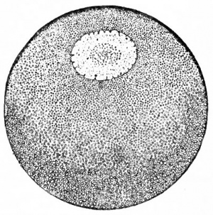
Most students have seen the eggs of the frog either in the laboratory or in a pond during the springtime. They probably have observed the little objects embedded in the jelly-like mass, scores of them in a cluster, each egg in its own gelatinous capsule, and all the capsules clinging to one another. Each ovum is a sphere, a little more than a millimeter in diameter in the common wood frog and as much as 3 mm. in some other species, with a dark side and a light side; and if the ovum has been at rest in its natural environment for a few minutes the dark side is uppermost (Fig. 26).
The dark color is due to the presence of brown pigment granules. The portion of the egg where there is less pigment contains an abundance of yolk globules suspended in the cytoplasm, while the darker part consists of cytoplasm with fewer yolk globules. The nucleus of the cell is located in the part containing the more cytoplasm and is therefore eccentric. The distribution of cytoplasm, yolk and pigment is apparently an expression of the internal organization of the egg, yielding here a visible polarity. The cytoplasmic or animal pole contains the nucleus and abundant pigment, the latter mostly near the surface; the yolk or vegetal pole contains less cytoplasm and pigment but abundant deutoplasm (Fig. 26). As far as determined, the egg is radially symmetrical around the axis extending from the center of the animal pole to the center of the vegetal pole; that is, assuming this axis to be vertical, the egg possesses the same organization in all radii drawn from the axis in any given horizontal plane. The polarity and symmetry _of the egg are important factors in development.
The eggs are expelled by the female frog into the water and the spermatozoa discharged by the male mingle with the egg clusters. A sperm burrows through the gelatinous capsule and thin vitelline membrane of an egg and enters the cytoplasm usually about 40 degrees from the center of the animal pole. There seems to be some determining factor in the entrance of the sperm at or near that particular parallel, but the point of entrance may lie in any meridian of the egg. The first sperm that enters the cytoplasm seems to set up changes, probably of a physico-chemical nature, which bar admittance to other sperms. The sperm head and the body containing the centrosome move through the cytoplasm for some distance toward the center of the egg, then rotate so that the body is in advance of the head and change their course in the direction of the egg nucleus. The trail of the sperm is marked by an extra amount of pigment, indicating probably some increase in cytoplasmic activity. The course of the sperm toward the center of the egg is the penetration path, the course toward the egg nucleus, the copulation path.
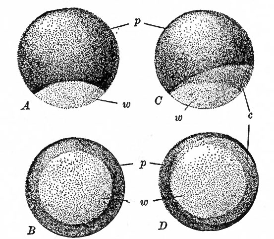
Fig. 27. A frog's egg before and after fertilization, showing the formation of the gray crescent. A, Unfertilized egg seen from the side; B, unfertilized egg seen from the vegetal pole. C, fertilized egg seen from the side; D, from the vegetal pole, c, Gray crescent; w, nonpigmented vegetal pole. Kellicott.
The sperm nucleus, as soon as it enters the egg, appears to stimulate the cytoplasm to activities leading to a rearrangement of the egg substances and thus to a reorganization. Beginning at the point where the sperm enters, the cytoplasm streams toward the animal pole and the yolk toward the vegetal pole, a sharper polar differentiation thus resulting. On the supposition that this influence of the sperm spreads like a wave from the point of entrance, it follows that the original rotatory symmetry of the egg is disturbed and a new symmetry established which is a bilateral one, with the plane containing the penetration path as the median plane. In other words the egg has become bilaterally symmetrical, with the plane of symmetry cutting the center of the animal pole, the center of the vegetal pole and the point of entrance of the sperm. There is also a visible external change in the distribution of pigment. On the side of the egg opposite the point where the sperm entered, some of the pigment granules over a crescent-shaped area at the lower border of the pigmented surface are carried from their original position, leaving this area lighter in color. The name, gray crescent, is given to the lighter area which extends more than half way round the egg (Fig. 27).
The rearrangement of the egg substances disturbs the center of gravity of the egg. The original axis, extending from the center of the animal pole to the center of the vegetal pole, is inclined at an angle of about 30 degrees to the vertical, the margin of the highly pigmented pole being tilted accordingly out of the horizontal. The gray crescent lies on the higher side. The vertical axis of the egg is now the gravitational axis, and, from the manner in which the internal rearrangement of egg substances has presumably occurred, a gravitational plane will bisect the egg into symmetrical halves, bisecting the gray crescent and containing both the gravitational axis and the original polar axis. All these changes have been caused or at least initiated by the sperm.
Cleavage
When the sperm nucleus reaches the egg nucleus via the copulation path the two nuclei join to form the single nucleus of the fertilized ovum. The sperm centrosome divides into two which take positions at opposite poles of the single nucleus. A spindle develops between the centrosomes, and the chromosomes assemble in the equatorial plane of the spindle. The direction that the spindle assumes does not appear to be wholly a matter of chance. In the first place it forms at right angles to the egg axis; for it is generally true that the spindle of a cell in division lies in the direction of the greatest cytoplasmic mass. If the egg is not subjected to pressure, the spindle tends to lie in the plane of egg symmetry or at right angles to it, although there may be many variations. If there is pressure from without, the spindle tends to lie at right angles to the direction of pressure. The factors other than pressure which influence the direction of the spindle have not been determined; but it appears that the spindle has a tendency at least, to assume a position of symmetry relative to the structure or internal organization of the egg. This means therefore that the first cleavage plane, which of course cuts the spindle at right angles, tends to divide the egg in or near the plane of symmetry or at right angles to it. In about 25 per cent, of instances the first cleavage plane deviates but little from the plane of egg symmetry; in about 10 per cent, it lies transversely to the plane of symmetry. It is also true that the first cleavage plane tends to coincide with the median plane of the future embryo. Summing up, it may be stated that there is a tendency in the frog for the median plane of the egg, the first cleavage plane and the median plane of the embryo to coincide; but, remembering that all these planes contain the egg axis, any other relation may be encountered.
On the surface the first cleavage furrow appears as a shallow groove on the pigmented side of the egg and then gradually extends around to the yolk pole. This is the surface indication of the division which separates the egg into halves or blastomeres. If the cleavage plane coincides with the plane of symmetry, the two blastomeres are symmetrical and the gray crescent is divided symmetrically; otherwise the two blastomeres are asymmetrical in internal structure. The division is total, but the two cells remain flattened against each other in close contact. It should be noted also that the division is retarded in the vegetal portion of the egg by the yolk globules in the cytoplasm. The retardation is so marked that the cleavage furrow of the second division appears at the animal pole before the first furrow has reached the vegetal pole. The second furrow crosses the first at right angles at the pigmented pole and extends around to the yolk pole in the same manner as the first. The second cleavage plane, of which this second furrow is the surface marking, intersects the first at right angles and thus divides each of the first two blastomeres into equal parts. The direction of the plane is determined by the position of the spindle in each primary blastomere, this lying in the direction of the greatest cytoplasmic mass. The first four blastomeres are approximately equal in size and contain equal amounts of cytoplasm and yolk. They remain in close contact so that collectively they still form a sphere which is marked on the surface by shallow grooves.
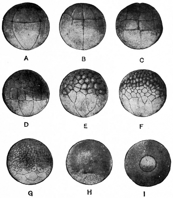
The third cleavage planes intersect the first two at right angles but lie nearer the animal pole than the vegetal pole, the furrow on the surface appearing about 60 degrees from the animal pole. In this manner the four blastomeres are divided into eight (Fig. 28, A). The upper four members are smaller and contain an excess of cytoplasm while the lower four are larger and contain an excess of yolk. This condition gives rise to the terms micromeres and macromeres. In some instances the third cleavage plane deviates from the latitudinal, even to being meridional, in one or more blastomeres. Typically the fourth cleavages are meridional, producing eight micromeres and the same number of macromeres. Here again the planes may deviate from the meridional position and disturb the typical pattern. Not all the blastomeres necessarily divide at the same time, as might be implied from the description. The lack of synchronism is especially true between micromeres and macromeres because in the latter the process of division is retarded to a marked degree by the inert yolk. From the fifth cleavage on, the micromeres very noticeably divide more rapidly than the macromeres with the result that the former become more numerous than the latter (Fig. 28, B, C, D, E, F, G). It is often stated that the rate of cleavage is directly proportional to the amount of cytoplasm and inversely proportional to the amount of yolk.
Fig. 28. Cleavage of the frog's egg. Morgan. A, Eight-cell stage; B, beginning of sixteen-cell stage; C, thirty-two-cell stage; D, forty-eightcell stage (more regular than usual); E, F, G, later stages; H, I, formation of blastopore.
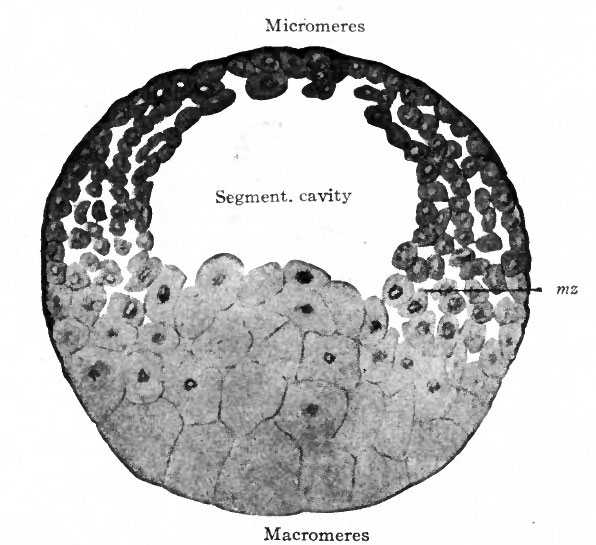
Returning for a moment to the first four blastomeres, the inner edge of each does not quite make contact with its neighbor, and so a minute space is left where the first two cleavage planes intersect. This rounding of the corners is probably due to the tendency for each cell to assume spherical form, which is the natural consequence of its semifluid nature and surface tension. When the third cleavage planes cut the first two at right angles somewhat above the equator, producing eight cells, the inner corners of these are rounded off and the space here is somewhat augmented. In the interior of the mass there is therefore a small cavity which, since the upper four cells are smaller than the lower, is eccentric. As the blastomeres continue to divide around it, the cavity increases in size but remains eccentric. During the first few divisions there is only a single layer of cells around the cavity; then some of the cells divide parallel to the surface and a double layer appears and then several layers. The multiplicity of layers is especially characteristic of the yolk cells. The entire structure is a hollow sphere called the blastula and the eccentric cavity within, known as the blastoccel or segmentation cavity, has a dome-shaped roof of micromeres and a floor of macromeres (Fig. 29). The peripheral stratum of closely compacted cells is the most highly pigmented while the cells beneath are less pigmented and somewhat more loosely arranged. The blastula is about the same size as the egg before it began to divide. It is similar to Amphioxus in that it is a hollow sphere, but is different in that the blastoccel is eccentric and the cells form several layers instead of one. (Compare Figs. 20 and 29.) As the cells multiply, those in the highest part of the dome-like roof of the blastoccel migrate toward the equator so that the roof becomes thinner and the lateral wall becomes thicker. The thicker lateral wall, which also exhibits rapid cell proliferation, is called the germ ring and probably corresponds to a similar zone of rapidly dividing cells in Amphioxus at the beginning of gastrulation. On the side of the blastula where the gray crescent is situated the germ ring migrates across the equator and down about halfway to the yolk pole. This downward migration displaces the yolk cells in the interior upward, producing an elevation in the floor of the blastoccel. As subsequent development proves, the side where the germ ring reaches the lowest point marks the caudal end of the embryo. During the formation and early migration of the germ ring the blastula increases about one-fifth in size but remains spherical. Some water perhaps filters into the blastoccel, although part of its contents is probably products of cell activities.
Fig. 29. From a sagittal section through blastula of frog. Bonnet, mz., Marginal zone.
Gastrulation
In the frog as in Amphioxus gastrulation comprises the change of a single-layered structure, the blastula, into a double-layered structure, the gastrula. The processes by which this change is effected are more complex in the frog, the visible factor in the complexity being the greater quantity of yolk. The inert yolk stored within an egg is always an influence in development.
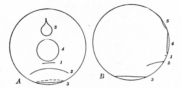
Viewing first the exterior of the blastula, a slight groove appears on the posterior side across the median sagittal plane at the lowest part of the germ ring, that is, about midway between the equator and the center of the yolk pole. The small pigmented cells bound the groove above, the larger yolk cells below (Fig. 28, H). As development proceeds the groove becomes longer, following the boundary between the two types of cells, which is of course the lower margin of the germ ring. It thus takes on the from of a crescent. Continuing to elongate in the same directon, the two horns of the crescent would eventually meet and the groove would thus become a ring encircling the blastula at the boundary between the pigmented and yolk areas. This actually occurs, but in the meantime the pigmented area extends farther down owing to the descent of the germ ring and the downward progress is more rapid on the posterior side where the groove first appeared. The result of this is that by the time the horns of the crescent meet to form a ring, the ring is much smaller than if there had been no downward movement; and since the original groove was bounded above by pigmented cells it now follows that the ring is bounded all round on the outside by pigmented cells. For the same reason the ring is bounded on the inside by yolk cells. These are the only yolk cells now visible on the surface. Subsequently the ring becomes still smaller and then flattened from side to side and finally reduced to a small slit. (See Fig. 30.)
The changes on the surface are merely partial expressions of the complicated processes in the interior. In a sagittal section of the blastula at the time the superficial groove appears, the initial step in these processes can be observed. The groove appears as a slight indentation above which are the smaller cells of the germ ring and below, the larger yolk cells. At this side is seen also the elevation of the floor of the blastoccel caused by the rising of the yolk cells; and there is a slight separation of this elevation from the smaller cells. The groove represents the beginning of a process of invagination which, however, is much less conspicuous than that in Amphioxus where the whole side of the blastula is invaginated. In the frog the yolk cells, laden with inert substance, are much less yielding to such factors as would produce invagination.
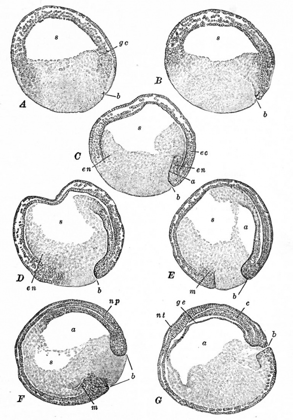
The successive stages of gastrulation as seen in sagittal section can be followed clearly in Fig. 31. The pictures are more vivid than verbal description. The groove can be seen to grow deeper in successive stages, turning upward into the elevation of yolk cells, seeming to push that elevation before it, and following the roof of the blastoccel across to the opposite side. When well on its way, the groove expands into a broad space which finally occupies the interior of the structure in much the same way as did the blastocoel. This broad space is the archenteron which opens to the exterior through the annular groove which was described on surface view, the opening being the blastopore. The yolk cells which are inside the ring can here be seen to fill the blastopore like a plug; collectively they are called the yolk plug. It should also be noted that the yolk cells form an elevation in the floor of the blastoccel on the side opposite the invagination. As a matter of fact the elevation occurs all the way round the blastoccel as does also the cleft between the elevation and the smaller cells.
Fig. 30. Diagrams showing the position of the blastopore at successive stages of gastrulation in the frog's egg. A, posterior view; B, lateral view. Figures 1-5 indicate the shape and position of the blastopore during the internal changes; figure 5 indicates its position after the rotation of the gastrula. Compare Figs. 31 and 35. Kellicott.
Invagination is probably not as important a factor here as it seems to be although it plays a part; it certainly is not as important as in Amphioxus. It must be remembered that the cells of the germ ring are multiplying rapidly when the invagination groove appears. The rapid proliferation continues during the processes thus far observed and many cells migrate inward around the lip of the blastopore. This perhaps is comparable with a similar series of rapid divisions and migration in the germ ring in Amphioxus. Consequently many of the cells that form the roof of the archenteron are not brought in by the invagination but by involution.
Fig. 31. Median sagittal sections showing successive stages of gastrulation in the frog's egg. Bracket, from Kellicott.
- A, beginning of gastrulation; B, slight advance in invagination and beginning of epiboly; C, invagination and epiboly progressing, inflection of cells (involution) occurring around dorsal lip of blastopore which is now an obvious structure; D, epiboly has resulted in covering of a large part of yolk by lip of blastopore; E, blastopore is now circular and filled with the yolk plug (cf. Fig. 30, A, 4) and the archenteron appears as a small space; F, the blastoccel is nearly obliterated; G, gastrulation completed.
- a, Archenteron; b, blastopore; c, rudiment of notocord; ec, ectoderm; en, entoderm; gc, gastrular cleavage, ge, entoderm (protentoderm) ; m, peristomal mesoderm; np, neural plate; w/, transverse neural ridge; s, blastocoel.
There is still another factor in gastrulation. It has already been noted that on surface view the groove moves downward as the highly pigmen ted cells along its upper or dorsal lip encroach upon the non-pigmented area, so that when the groove becomes ring-shaped only a small yolk area is visible. This downward growth over the yolk area, or epiboly, which is more rapid on the side where the groove began, results in the enclosure of more and more yolk cells so that only those comprising the yolk plug are left exposed. It is this process (epiboly) therefore which causes the lessening of the crescent and ring as seen on surface view. (Compare Fig. 30.)
These processes which are grouped under the term gastrulation have converted the single-layered blastula into the double-layered gastrula. The outer layer composed of several strata of pigmented cells is the ectoderm which is in contact with the environment. The inner is the entoderm which lines the archenteric cavity. Two types of entodermal cells are distinguishable: those forming the roof and sides of the archenteron which contain a moderate amount of pigment and those forming the floor which hold little pigment but an abundance of yolk. The two primary germ layers are continuous at the rim of the blastopore.
Two other features which are incidental to the processes of gastrulation must be noted because of their bearing upon future development. Recalling the migration of the crescentic groove, which eventually becomes the ring around the yolk plug, it is obvious from the manner in which the migration occurs that the cells along the horns of the crescent are drawn toward the median region. The name given to this phenomenon is concrescence. The result of it is that the cells are piled up in a median linear strand, from which the rudiments of certain organs emerge. The outer feature is the flattening of the ring from side to side, concomitant with the withdrawal /inward and disappearance of the yolk plug, so that the two lateral margins approximate, leaving only a narrow slit leading from the exterior into the archenteric cavity. Subsequently the slit is closed by fusion of its walls, but part of the depression in its site becomes the anal pit or proctodaeum.
At this stage the gastrula is still spherical and only slightly larger than the blastula. It possesses the same fundamental arrangement of structure as the gastrula of Amphioxus. The ectoderm forms contact with the environment, implying response to stimuli and protection; and the organs correlated with these functions are derived from this layer. The archenteric cavity with its lining of endoderm is confined to the interior of the developing organism and comprises the primitive alimentary system. Within the cells of the entoderm is the food that must suffice until the animal reaches a stage when it is able to obtain a supply from the outside; but the rudiment of the future complex alimentary mechanism is already formed. The blastopore is not a free opening, as in Amphioxus, but is obstructed by the yolk plug. The latter is eventually withdrawn and the anus develops in the site of a part of the blastopore. The mouth is a new opening which develops at the forward end of the gut. A somewhat more detailed discussion of the biological significance of the blastula is given on page 40, in the chapter on Amphioxus.
Mesoderm Formation
In order to detect the beginning of the middle germ layer it is necessary to look back into the period of gastrulation. Gastrulation and mesoderm formation overlap each other. In a sagittal section of the blastula just as gastrulation commences, the cells of the germ ring are continuous with the yolk cells above the groove that indicates the beginning of in vagina tion (Fig. 31, A). This transition zone, traced through the subsequent stages of development, is composed of cells which occupy a position always in the angle between ectoderm and entoderm and merge with these layers (Fig. 31, B, C, D, E, F). The cells in question comprise the early mesoderm. Appearing as it does in the angle between the other layers in the lip of the blastopore, it is obvious that when the blastopore becomes circular the mesoderm takes the form of a circular band. In Amphioxus it was clear that the mesoderm originated from entoderm (see p. 42), but in the frog the first mesodermal cells bear such relation to the other layers that their origin is not so readily determined. In later stages, however, it will be apparent that mesoderm arises from yolk entoderm.
In the description of gastrulation it was pointed out (p. 58) that during the migration of the crescentic groove and its transformation into a ring the cells along the horns of the crescent were drawn medially and piled up in an axial strand which then extended upward and forward from the dorsal lip of the blastopore. The mesodermal cells appear in the dorsal lip of the crescentic groove and, as the migration of the groove goes on, they are affected in the same way as the other cells in this region. Therefore the band of mesodermal cells around the blastopore is broader at the dorsal side. In other words, a band of mesodermal cells extends upward and forward from the dorsal lip of the blastopore, forming a part of the axial strand. And since the proliferation and involution of cells, which occur during gastrulation, tend to carry the mesodermal cells upward and forward and since the mesodermal cells themselves are proliferating, the mesoderm soon becomes almost as extensive dorsally as the entoderm.
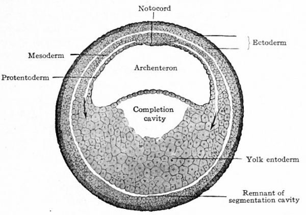
Fig. 32. Transverse section of embryo of frog (Rana fusca). Bonnet. The section is taken in front of (anterior to) the blastopore.
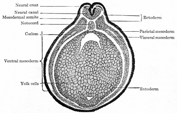
Fig. 33. Transverse section through embryo of frog (Rana fusca). Bonnet.
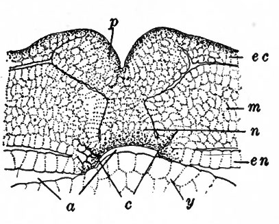
In the dorsal axial strand of cells, which later will be considered more in detail, the three layers are at first merged. Lateral to this the mesoderm becomes clearly delimited from ectoderm, at least a potential cleft separating the two layers. For a short distance laterally the mesoderm also becomes delimited from entoderm, but farther laterally it is fused with entoderm (Fig. 32). Then as development proceeds the superficial cells of the yolk entoderm, with which the mesoderm is merged, become differentiated and split off or delaminated and added to the mesoderm. In this manner the mesoderm becomes more extensive until finally it reaches all the way round ventrally between the other layers, although it is not complete for some time (Fig. 33). There is ample evidence here that this portion of the mesoderm is a derivative of entoderm (yolk entoderm). The mesoderm that develops along the crescentic groove and around the blastopore is often called peristomal; that which arises elsewhere is known as gastral mesoderm. The behavior of the mesoderm that is involved in the dorsal axial strand above or anterior to the blastopore is rather complex because out of that strand arises one of the early axial structures of the embryo, the notocord. First a slight cleft between ectoderm and mesoderm gradually extends from each side toward the mid-dorsal line, but just before reaching the line abruptly turns ventrally. This cleft as it bends ventrally leaves a group of cells in the axial line which is still continuous with ectoderm above and entoderm below. The axial group of cells is the rudiment of the notocord. (Fig. 34) Just above or anterior to the blastopore, in the region where entoderm and mesoderm are still continuous with at the lower lateral angles of the notochord rudiment, a pair of grooves appear which are not particularly conspicuous but which seem to be evaginations from the archenteron (Fig. 34).
Fig. 34. Portion of a transverse section still continuous at the lower lateral angles of the larva of a frog (Rana fusca). Hertwig. a, Archenteron; c indicates enterocoel formation; ec, ectoderm; en, entoderm; m, mesoderm; , notocord; p, neural plate; y, yolk entoderm.
Farther forward the grooves are slightly more conspicuous, but still farther forward disappear. It has been argued that these grooves are homologous with the enteroccelic evaginations in Amphioxus where the mesoderm arises by outgrowth from the entoderm. On the other hand it has also been argued that the more primitive mode of mesodermal development is represented in the frog and that the simplicity of origin is secondarily acquired in Amphioxus. Whether pr not these grooves are enteroccelic evaginations in the frog, soon after their appearance the notocord rudiment becomes separated from entoderm below, from ectoderm above, and lies in the axial line between what are now the paraxial masses of mesoderm on the two sides (Fig. 33) The notocord therefore becomes an independent structure except at its caudal end where it merges with all the layers which in turn are merged with one another at the blastopore. A similar fusion is present in Amphioxus (p. 45) ; and out of this mass of cells, as development proceeds from before backward, the three germ layers and the notocord are differentiated.
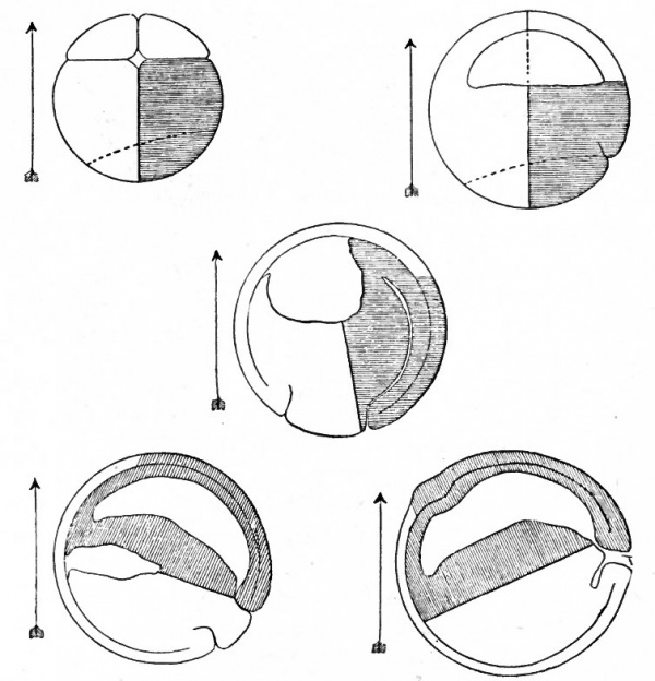
During gastrulation a certain shift in position of the structure as a whole is observed. In the completed blastula it was noted that the yolk pole was directed downward owing to the slightly higher specific gravity of the yolk. During gastrulation it is obvious that the center of gravity of the whole mass is shifted. This can readily be seen if one follows the changes in sagittal sections (Fig. 31). As a result the whole structure rotates through an angle of about 90 degrees (Fig. 35). The blastopore therefore assumes a position which is nearly on a level with the center of the gastrula. After the rotation the dorsal and ventral sides and the cephalic and caudal sides (ends) of the gastrula and of the future embryo are fixed.
Fig. 35. Diagrams of median sagittal sections through an eight-cell stage and four stages during gastrulation of the frog's egg. Kopsch, from Kellicott. The arrow marks the vertical. If one compares the shaded parts of the figure with the fixed vertical line it is seen that the gastrula rotates through an angle of about 90 degrees in the counter-clockwise direction.
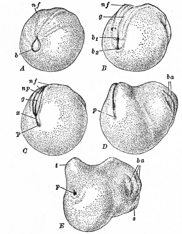
The completed gastrula is still spherical, but then at once it begins to elongate in the direction of the axis drawn from the blastopore to the opposite pole. The dorso-caudal region is drawn out into a bud-like structure; the dorsal side becomes flat or slightly concave in the cephalo-caudal direction; the ventral side remains broadly convex owing to the presence of the yolk in the ventral wall of the archenteron (gut) (Fig. 36). This growth in length is the beginning of the characteristic cephalo-caudal elongation of the larva and adult. It is due in part to proliferation of cells generally but chiefly due to proliferation at the caudal end in the region dorsal to the blastopore. Here as in Amphioxus growth takes place largely from before backward; and the bud-like process in the dorso-caudal region is one of the outward expressions of the growth.
Fig. 36. Postero-lateral views of successive stages following gastrulation in the frog. Ziegler, from Kellicott.
- A, blastopore in process of closing, neural folds slightly indicated; B, gastrula slightly elongated, blastopore closed, neural groove and folds obvious; C, anal portion of blastopore still visible at bottom of proctodaeum, neural folds closing dorsally; D, neural folds nearly closed, branchial arches appearing, tail bud forming; E, neural folds fused, tail bud more conspicuous.
- b, Blastopore containing yolk plug; bi, dorsal part of blastopore (rudiment of neurenteric canal) ; fe, ventral part of 'blastopore (rudiment of anus); ba, branchial arches; g, neural gro ve ; nf, neural folds; np, neural plate; p, proctodaeum, with anal portion of the blastopore at the bottom; s, oral sucker; t, tail bud; x, neural folds covering the blastopore thus establishing the neurenteric canal.
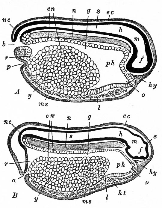
In order to bring the development of the frog up to a point corresponding to the stage of Amphioxus reached at the end of the previous chapter, it is necessary still to consider briefly the appearance of the neural tube and some further changes in the mesoderm. During the latter part of the gastrulation period a band of ectoderm extending forward from the dorsal lip of the blastopore over the dorsum of the gastrula becomes slightly thicker. This band of cells, the neural plate, is narrow near the blastopore and becomes broader farther forward (Fig. 36,5). During the withdrawal of the yolk plug and the closure of the blastopore the margin of the plate becomes thicker and elevated above the surface level to form the neural ridges. The depression between the ridges is the neural groove (Fig. 33). At the cephalic end of the plate the ridges curve medially and meet each other, forming the transverse neural fold. The ridges grow higher, the groove becoming correspondingly deeper, and finally lean far enough toward the median sagittal plane to meet and fuse in the mid-dorsal line, so that a tube is formed with the lumen as the central canal. The fusion of the ridges usually begins about midway between the cephalic and the caudal ends, and then continues forward and backwards. The caudal portion of the neural tube encloses the dorsal part of the blastopore which thus, as in Amphioxus, becomes the neurenteric canal, the communicating aperture between the central canal and the archenteron (Fig. 37). After the dorsal closure of the tube the non-neural ectoderm forms a continuous layer so that the tube is completely covered (Fig. 33). The broader cephalic portion of the neural tube is the rudiment of the brain, the narrower remaining part is the beginning of the spinal cord. In the mesoderm lateral to the neural plate and notocord the cells for a time proliferate more rapidly than elsewhere and produce a rather stout mass (Fig. 33) extending from the head to the blastopore. From this mass laterally the mesoderm extends between ectoderm and entoderm as previously described. Just behind the head region the paraxial mass suffers a rearrangement of its cells so that a block is delimited transversely. Just behind this another block is formed in the same manner; a similar process produces a third, and so on toward the caudal region. The blocks themselves consist of closely compacted cells while in the intervals between them the cells are loosely arranged. These blocks are the mesodermal somites which in their arrangement express the fundamental metameric or segmental principle of all vertebrates and many invertebrates. Lateral to the somites a cleft appears in the originally single layer of mesoderm thus dividing it into two layers (Fig. 33). The cleft commences near the somites and gradually extends all the way round ventrally; it also extends into some of the somites. This cleft is the rudiment of the ccelom or body cavity, and its extension into the somites probably corresponds to the myoccel in Amphioxus The layer of mesoderm ectal to the coelom and apposed to the ectoderm is called the somatic or parietal layer; the layer ental to the ccelom and apposed to entoderm is the visceral or splanchnic mesoderm.
Fig. 37. Median sagittal sections of frog larvae. Marshall. A , just prior to closure of blastopore ; B, just after closure of blastopore. a, anal aperture; b, blastopore; e, epiphysis; ec, ectoderm; en, entoderm; f, fore-brain; g, mid-gut; h, hind-brain; ht, rudiment of heart; hy, hypophysis; I, rudiment of liver; m, mid-brain; ms, mesoderm; n, no tocord; nc, neurenteric canal ; o, oral evagination; p proctodaeum; ph i, pharynx; r, rectum; s, spinal cord; y, yolk entoderm.
At this stage of development in the frog the fundamental vertebrate organization is expressed in the general arrangement of structure (Fig. 37). The body as a whole consists of a tube within a tube; the rudimentary digestive system, extending lengthwise in the growing animal, is the inner tube, the outer tube is the body wall, and the body cavity or ccelom separates the two tubes. The dorsally located neural canal, the notocord around which the vertebral column develops in the true vertebrate, and the series of mesodermal somites are at this time simple structural patterns from which the complex vertebrate organization is evolved.
- Next: Chick
References for Further Study
BRACKET, A.: Recherches sur Fontogenese des Amphibiens urodeles et anoures. Archives de Biologie, tome 19, 1902.
EYCLESHYMER, A. C.: The early Development of Amblystoma, with Observations on some other Vertebrates. Journal of Morphology, Vol. 10, 1895.
HERTWIG, R.: Furchungsprozess. In Hertwig's Handbuch der vergleichenden und experimentellen Entwickelungslehre der Wirbeltiere. Bd. I, Teil I, Kap. Ill, 1903. Contains extensive bibliography.
JENKINSON, J. W.: On the Relation between the Symmetry of the Egg and the Symmetry of Segmentation and the Symmetry of the Embryo in the Frog. Biometrika, Vol. 7, 1909.
KELLICOTT, W. E.: Chordate Development. Chap. II, 1913.
MORGAN, T. H.: The Development of the Frog's Egg. 1897.
| Historic Disclaimer - information about historic embryology pages |
|---|
| Pages where the terms "Historic" (textbooks, papers, people, recommendations) appear on this site, and sections within pages where this disclaimer appears, indicate that the content and scientific understanding are specific to the time of publication. This means that while some scientific descriptions are still accurate, the terminology and interpretation of the developmental mechanisms reflect the understanding at the time of original publication and those of the preceding periods, these terms, interpretations and recommendations may not reflect our current scientific understanding. (More? Embryology History | Historic Embryology Papers) |
Text-Book of Embryology: Germ cells | Maturation | Fertilization | Amphioxus | Frog | Chick | Mammalian | External body form | Connective tissues and skeletal | Vascular | Muscular | Alimentary tube and organs | Respiratory | Coelom, Diaphragm and Mesenteries | Urogenital | Integumentary | Nervous System | Special Sense | Foetal Membranes | Teratogenesis | Figures
Glossary Links
- Glossary: A | B | C | D | E | F | G | H | I | J | K | L | M | N | O | P | Q | R | S | T | U | V | W | X | Y | Z | Numbers | Symbols | Term Link
Cite this page: Hill, M.A. (2024, April 27) Embryology Book - Text-Book of Embryology 5. Retrieved from https://embryology.med.unsw.edu.au/embryology/index.php/Book_-_Text-Book_of_Embryology_5
- © Dr Mark Hill 2024, UNSW Embryology ISBN: 978 0 7334 2609 4 - UNSW CRICOS Provider Code No. 00098G
