Book - Manual of Human Embryology 4
Keibel F. IV. Young human ova and embryos up to the formation op the first primitive segment in Keibel F. and Mall FP. Manual of Human Embryology I. (1910) J. B. Lippincott Company, Philadelphia.
| Historic Disclaimer - information about historic embryology pages |
|---|
| Pages where the terms "Historic" (textbooks, papers, people, recommendations) appear on this site, and sections within pages where this disclaimer appears, indicate that the content and scientific understanding are specific to the time of publication. This means that while some scientific descriptions are still accurate, the terminology and interpretation of the developmental mechanisms reflect the understanding at the time of original publication and those of the preceding periods, these terms, interpretations and recommendations may not reflect our current scientific understanding. (More? Embryology History | Historic Embryology Papers) |
IV. Young Human Ova and Embryos up to the Formation of the First Primitive Segment
(a critical. account)
By Franz Keibel, Freiburg i. Br.
By the term ovum is understood in human embryology not only the egg-cell but also later the entire structure developed from the egg-cell, the embryo or fetus surrounded by the amnion and chorion. In this sense the word is used here. I do not intend to enumerate and describe here all young and very young human ova that have been observed, but only those which may be regarded as normal or approximately so, and as such I can regard only those in which an embryo has been observed. The observations of Graf Spee and Peters on human ova, and of Selenka on those of monkeys, have shown that in man and the primates the chorion grows much more rapidly than the embryo, that, consequently, a relatively large ovum may contain a very small embryo, and that even in the youngest ova yet studied the amnion and the yolk sack are already formed. I shall show later that in all probability the embryo never lies free upon the surface of the ovum, as it does in the birds and in many mammals, but that from the beginning it is sunk in the interior of the ovum, and that the amniotic cavity arises as a cleft and not by the formation of folds, and is always closed. The extraordinary minuteness of the embryonic anlage is a sufficient explanation why in early times, when the methods of investigation were imperfect, it was overlooked or unrecognized ; many of these earlier described ova may have been normal or nearly so. But when no embryonic anlage is found in an ovum that has been investigated according to all the rules of modern technic, as is the case with that which G. Leopold[1]has lately studied with so much care, that ovum is certainly to be regarded as pathological; and the occurrence of maternal blood in the interior of the ovum is further evidence in this direction, as Spee has pointed out in Schwalbe's Jahresbericht. Leopold's ovum does not, therefore, require consideration here ; on the other hand, some older observations may be noticed, such as those of Eeichert, Wharton Jones, and Breuss. The ovum of Reichert especially has played and is still playing, though improperly, an important role in human embryology.
Reicher[2] found the ovum in the uterus of a suicide and estimated its age at twelve to thirteen or thirteen to fourteen days. It was completely enclosed by the mucous membrane of the uterus. On the side of the capsule which was turned towards the uterus there was a transparent area measuring 3 mm., which Reichert termed the capsule sear, believing that at the sides of it the mucous membrane of the uterus had grown up to surround the ovum. The ovum itself was a lenticular vesicle, whose diameters were 5.5 and 3.3 mm. The surface of the vesicle which was turned towards the uterus, the basal surface, was almost flat, that turned toward the lumen of the uterus somewhat curved. The marginal zone was richly furnished with small villi, the largest of which were 0.2 mm. in length and were already partly provided with lateral branches. From the margin small villi, diminishing in size, extended for some distance upon the surface of the vesicle turned toward the uterus wall (the basal surface of Reichert) ; but at the centre of the surface an area of about 2.5 mm. diameter remained free from them. In the centre of this free area Reichert described a dull circular spot. The surface of the ovum turned toward the lumen of the uterus was free from villi. The statements that Reichert makes concerning the finer structure of the ovum are in part insufficient and in part quite erroneous. Thus the wall of the ovum could not have been, as he supposed, purely epithelial, nor the villi hollow epithelial structures, but the wall must have been formed of mesodermal tissue with an epithelial covering and the axes of the villi occupied by mesodermal tissue. Reichert, indeed, perceived this mesodermal tissue, but regarded it as coagulated material. Also what he says concerning the ingrowth of the villi into the uterine glands is undoubtedly incorrect. As regards the structure of the dull spot on the uterine surface of the ovum, he supposed that it was formed by a layer of small, finely granular, nucleated, polyhedral cells, situated within the epithelial wall. It was taken for the embryonic anlage, and His has estimated the diameter of this "embryonic spot" as 1.6 mm. That this spot really represents the uninjured embryonic anlage is improbable, and Kollmann's statement in his " Handatlas der Entwicklungsgeschichte des Menschen" (1907) — "From what we know from the mammals this spot would now be regarded as the embryonic shield " — is, as will be shown later, absolutely disproved. A definite opinion cannot be given on account of the insufficiency of Reichert's description; nevertheless, I regard as well founded the conclusion of Spee, that Reichert had destroyed the actual embryonic structure during his preparation of the ovum and that in its degree of development it would have occupied a place between the oviun of Peters, to be described in detail later, and the Van Herff ovum of Spee. Probably Reichert had casually obsen^ed the embrj'o; it may have been the spherical body on the basal wall which he mentions on p. 26.
Another ovum which deserves mention is that described by Wharton Jones[3] in 1837; it was of the size of a pea. The fisrure drawn from the preparation in alcohol shows a diameter of 6.2 X 4.7 mm. The surface turned toward the lumen of the uterus was free from villi Imbedded in the cavity of the ovum was a spherical body, with a diameter of 1,5 mm., which His, probably correctly, identified as the embryonic structure, that is to say, the actual embryonic anlage together with the amnion, yolk sack, and belly stalk. His assumes that this embryonic structure may have been artificially displaced.
Next comes an ovimi described by Breuss[4] in 1877. It was expelled together with the entire lining of the uterus. The wall of the ovum, which had a diameter of 5 mm., consisted of two layers, the outer of which was epithelial and the inner formed of connective tissue. The villi were for the most part unbranched and were about 1 mm. in length and 0.07 mm. in diameter; they left free a roundish area 2 mm. in diameter. Vessels could not be distinguished in their interior. A projection which occurred in the interior of the ovum, consisting of nucleated cells and having a length of 1 mm. and a diameter of 0.5 mm., may have been the embiyonic structure. If it is assumed that the ovum was normal, we must suppose that Breuss overlooked the amniotic cavity and the cavity of the yolk sack, a supposition which I regard as possible.
Mention may also be made of two other ova, described by Allen Thomson.[5] In one of these the embryonic structure must be regarded either as pathological or as not corresponding to the degree of development of the oviun. Thomson estimates the age of the smaller of the two ova at twelve to thirteen days. It had a diameter of 6.6 mm., was everywhere surrounded with villi, and was almost completely filled by a vesicle which was apparently the yolk sack. Upon the yolk sack was an embryo 2.2 mm. long and with both its cranial and caudal ends separated from the sack. Thomson makes no mention of an amnion, but we may suppose that it was present and covered the embryo on the surface opposite the yolk sack; and a belly stalk must also have been present, since Thomson states that the embryo was attached by its dorsal surface to the external egg membrane, that is to say, to the chorion.
Although the ovum is larger than that of His, to be described below, the second observation of Thomson may be recorded here; it concerns an ovum measuring 13.2 mm. in diameter, whose age was estimated at fifteen days. It was oval in shape and was evenly surrounded with villi. In the interior was a large cavity filled with fluid; and at one spot was the embryo, closely attached to the chorion and with a yolk sack and the remains of the amnion. The embryo had a length of 2.2 mm. and its cranial and caudal ends projected somewhat beyond the yolk sack. Viewed from the surface turned toward the chorion, it showed distinctly the medullary folds, which manifested a tendency to fuse at the middle of their length; ventrally was the heart. The diameter of the yolk sack was also 2.2 mm.; nothing is said concerning an amnion, but a lobe which is shown in Thomson's figure at the head end of the embryo is apparently the remains of an amnion that had been destroyed during the preparation of the embryo. It is interesting to note that Kolliker[6]in 1879 regarded this second ovum described by Thomson as not quite normal on account of the large space which separated the embryo and yolk sack on the one side from the inner surface of the chorion on the other. We now know that this is the normal condition in embryos of this stage; and it is rather the smaller of Thomson's ova, which Kolliker was inclined to regard as normal, that shows abnormal conditions, since the yolk sack never fills the chorion so completely either in human or mammalian ova of this stage.
In all the ova so far mentioned a correct identification of the embryo, the yolk sack, amnion, belly stalk, and chorion is possible only in the two described by Thomson, which contained embryos already rather well developed; and even in these the identifications were only general ones, as may be seen from Kolliker's conmients upon the ova. He regarded, on the basis of the information available at that time, the normal ovum as abnormal and the abnormal one as normal, and the same conclusion was reached by Ecker, another distinguished embryologist of the time. A definite idea of the relations of the amnion and the belly stalk was also impossible for Kolliker. A correct interpretation of the discoveries mentioned could not be given at the time of their publication and, indeed, in part, not for some time after. The investigators who sought such interpretations were led from the right path, and Reichert's ovum, as I have stated, gave rise to many false ideas, even up to recent times. Consequently, as Elze and I[7] have already pointed out in our "Normentafel zur Entwicklungsgeschichte des Menschen," an observation by His[8] marks an important advance. The ovum in question (No. XLIV [Bff.] of His's collection) had a greater diameter of 8 mm. and, at right angles to this, a diameter of 7 mm.; it was somewhat flattened and at one point the villi were somewhat fewer than elsewhere. " On opening it there was found, on one wall, a small body measuring 1.4 mm. in its longest diameter and consisting of an ellipsoidal opaque body with a transparent vesicle attached to it. The opaque body, which seemed from partial foldings of its surface to be hollow, had a greater diameter of 0.85 mm. and a diameter at right angles to this of 0.6 mm. The transparent vesicle surrounded by its border one end of the ellipsoid. The connection with the chorion was by means of a short stalk, which stood in relation to both the vesicle and the ellipsoid." " I regard," His says in another place, " the more solid body as the umbilical vesicle and the transparent part as the amnion, and conclude from this that the embryonic anlage, so far as it is present, lies at the boundary between the two. With this idea the manner in which the structure is attached to the chorion agrees. That is to say, the place of attachment lies on the boundary between the vesicle and the opaque body." His's interpretation, we can say to-day with all certainty, is in agreement with the actual facts, and His was the first to give a perfectly correct interpretation of a human ovum of this stage. He further reported concerning this oviun that to the lower pole of the yolk sack there were attached threads of that looser tissue "which traverses the cavity of the ovum, one of these threads being especially distinguished by its tougher consistency and its opacity."
Later observations of young human embryos, carried out with the methods of modem technic, and especially with the aid of well stained and perfect series of sections, have, as has been already stated, confirmed His's views and have led to further, partly unexpected, results. It was the observations of Graf Spee, especially, that opened the way, and, later, H. Peters rendered great sendee; but for the sake of continuity the ova in question will not be described in the order in which they were discovered, but according to their degree of development. Consequently I shall begin with the ovum described by Bryce and Teacher.[9]
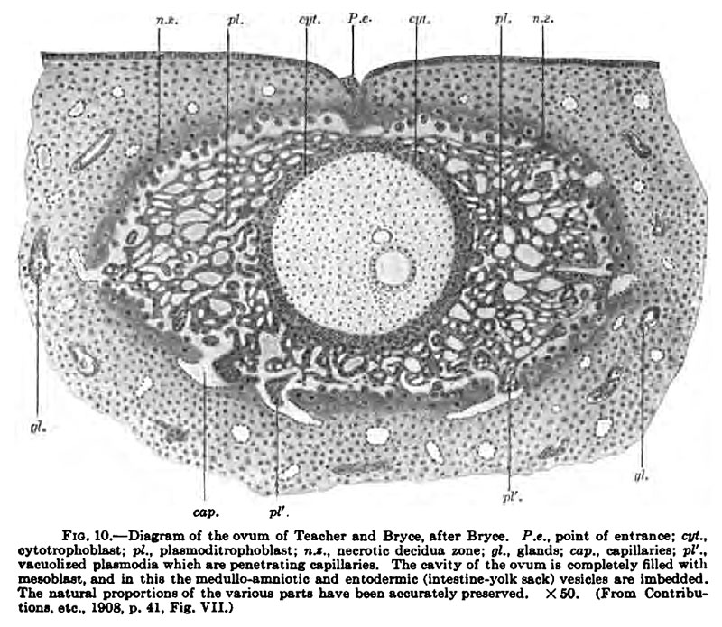
The ovum was obtained from an abortion, and although the presen'ation of the embryo proper is not perfect yet it is of the greatest importance; Fig. 10 shows the ovum as it lay in the uterine mucous membrane, according to a diagram by Bryce. With the exception of a small area it is completely surrounded by decidua, and the opening in the decidua (capsularis) is closed by coatated fibrin containing leucocytes. A large opening with fungoid tissue (the closing coagulum of Bonnet) is wanting.
The ovum, surrounded with blood, lies in a relativelj laige chamber, with whose walls it is not united; the maternal and fetal tissues are quite separate. The uinermost layer of the decidua, which forms the capsule of the ovum, is in an advanced stage of coagulation necrosis and, together with some deposits of fibrin, fonns around the ovum a capsule of dead tissue, which is interrupted only at one or two places, where blood-vessels open into the egg chamber, and at one where a hemorrhage has broken through into it.
Fig. 10. Diagram of the ovum of Teacher and Bryce. after Bryce.
The wall of the ovum consists of an inner layer (the cytotropboblast, the layer of Langhans's cells), whose cells are poorly separated from one another and externally pass over into a very irregnilarly arranged plasmoditropboblast ; tbis has a distinctly plasmodial character and forms a loose network, whose spaces are filled with maternal blood. The cytotropboblast is nowhere continued into the plasmoditropboblBBtic trabecule. The cavity of the ovum is oCoupied hj a delicate tissue which has the characters of mesenchyme. This primitive mesoblast shows no traces of a splitting into a parietal and a visceral layer; there is, accordingly, no Coelom. Alao, projections of the mesoblast toward the cytotropboblast (mesodermic villi) are not yet present.
The embryonic structures (the embryo together with the amnion and yolk sack) are represented by two vesicles which have an excentric position, and are completely separated from the cytotrophoblaat by the mesenchymstous tissue.
The cavity of the larger vesicle is supposed to be the amniotic cavity and that of the smaller the cavity of the yolk sack. The cells which enclose the amniotic cavity are cubical and those enclosing the yolk sack are flattened, but in neither vesicle do they show individual differentiation.
The egg-chamber is oval in form, the longest axis, lying parallel to the surface of the uterus, measuring 1.95 mm.; perpendicular to this and also parallel to the uterine surface the lumen of the egg-capsule measures 1.1 mm. and its depth (perpendicular to the surface of the uterus) is 0.95 mm. Since the wall of the ovum itself is folded the measurements of the cavity can be stated only approximately as 0.77 and 0.63 mm. The relative sizes of the amniotic cavity and of the yolk sack may be perceived from Fig. 10 ; unfortunately these structures were not intact.
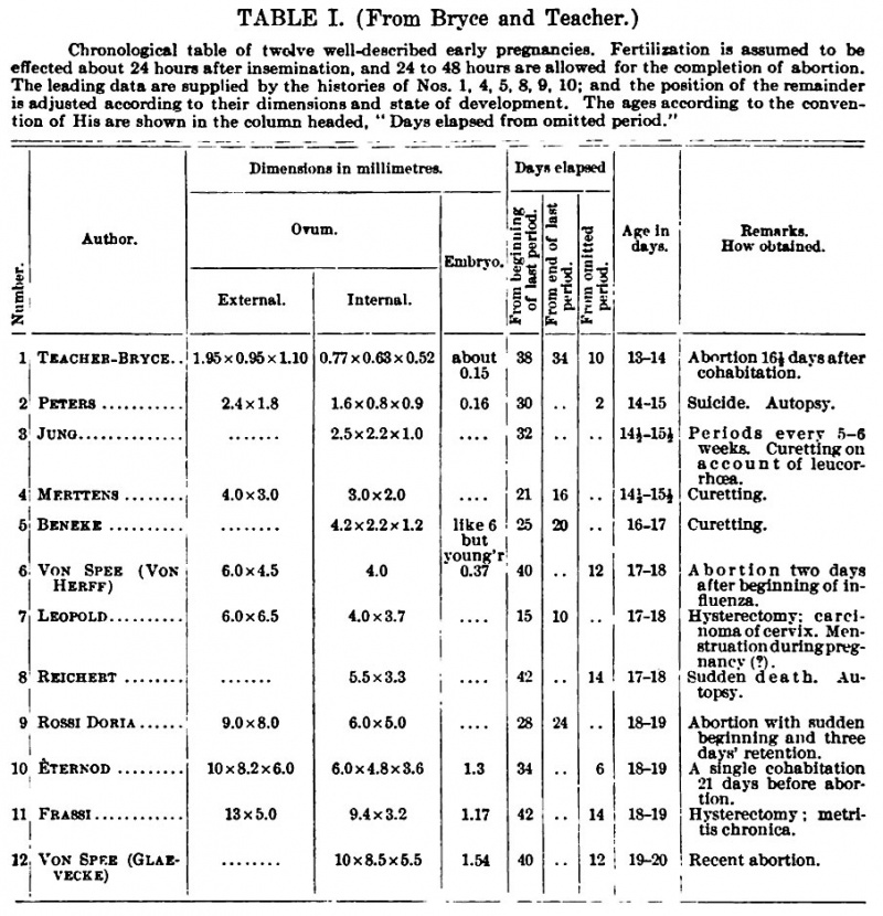
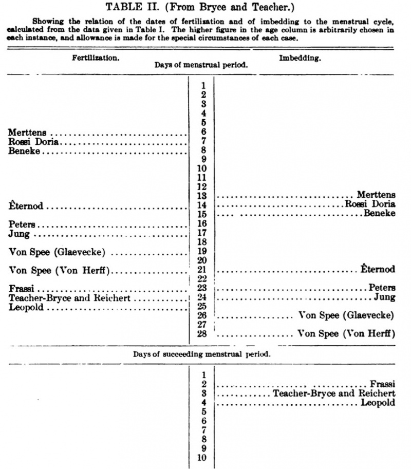
The estimate of the age of the ovum made by Teacher and Bryce is very interesting and important, since it is based on exact data concerning the menstruation and the cohabitations. They estimate the age at thirteen to fourteen days, the ovum haWng been expelled sixteen and one-half days after the fertilizuig coitus. The cause of the abortion is supposed to have been a later coitus. If this estimate be correct the other ova to be described later on are all older than has hitherto been supposed. The table given by Bryce and Teacher may be reproduced here.
Table I
(From Bryce and Teacher.)
Chronological table of twelve well-described early pregnancies. Fertilization is assumed to be effected about 24 hours after insemination, and 24 to 48 hours are allowed for the completion of abortion. The leading data are supplied by the histories of Nos. 1, 4, 6, 8, 9, 10; and the position of the remainder is adjusted according to their dimensions and state of development. The ages according to the convention of His are shown in the colunm headed, " Days elapsed from omitted period."
Table II
(From Bryce and Teacher.)
Showing the relation of the dates of fertilisation and of imbedding to the menstrual cycle, calculated from the data given in Table I. The higher figure in the age column is arbitrarily chosen in each instance, and allowance is made for the special circumstances of each case.
We now come to the ovum described by Hubert Peters[10]; it was obtained at the autopsy of a woman who had poisoned herself with caustic potash. Death had occurred within three hours after the taking of the poison and the autopsy was performed on the same day, a few hours after death. The ovum, together with the entire egg capsule, fixed by Prosector Kretz in Muller's fluid and hardened in alcohol, was successfully stained and microtomized by Hochstetter; it found in Peters an exceedingly careful observer. The uterus, from which the ovum was taken, was the size of a goose's egg and thick-walled, and felt somewhat more doughy and softer than a normal utenis. "The decidua of the corpus uteri was traversed by numerous furrows which crossed one another at various angles and occasionally formed grooves, so that the mucous membrane between these bounding furrows formed root-like or occasionally rounded projections toward the uterine lumen. In the middle of the posterior wall Prosector Kretz noticed a small area which was of the size of a hemp seed, which was somewhat paler but not prominent"; it contained the ovum. This was ellipsoidal in form, its diameters being 1.6 X ^-8 X ^-9 nam., these measurements, however, being of the cavity of the egg capsule. The part of the decidua in which the ovum lay presented, as the study of the sections showed, a slight, rounded elevation toward the cavity of the uterus. " While the decidua stretched as a very thin sheet, in the form of a capsulaxis, over the lateral portions of the ovum, the summit of the ovum was quite free from maternal tissue and projected freely into the lumen of the uterus by means of a blood-granulation mass which rested upon it " ; this mass Peters terms the fungoid tissue (Gewebspilz). If the interpretation which Peters gives of the fungoid tissue is correct and it is not a post-mortem phenomenon or a result of the poisoning, we have for the first time a human ovum that is not yet quite covered by a capsularis. Later Spee {Verh. Anat, Ges., 1902 (discussion of Marchand's paper) ; compare Schwalbe's Jahresber,, n. F., vol. viii. No. 2, p. 298) stated that occasionally young ova were to be found in which the egg capsule had an opening toward the uterus at the place where the scar tissue occurred, and at the corresponding place the ovum of Bryce and Teacher showed, as has been mentioned, only a small opening and no well-marked fungoid tissue. Upon the mesoblast layer of the ovum, which shows indications of the first few and as yet but slightly developed villi, follows a layer of epithelial cells, which reaches in places a thickness of more than 0.5 mm. and is traversed in a honeycomb manner by smaller and larger blood lacun®, but still remaining continuous at the periphery. Peters interprets this rightly, as I believe, as an " ectoblast shell " and names it the trophoblast, adopting the term which Hubrecht had proposed as a result of his observations on the hedgehog. Its further significance will be fully considered in the chapter on placentation and it will there also be compared with the corresponding structures of other mammals (cheiroptera) and with what is found in older human ova (Kastschenko, Merttens, Von Herff). As in the ovum of Bryce and Teacher differentiation of the cells of the trophoblast investment was evident, and the cytotrophoblast (Langhans's cells) and the spongiotrophoblast were distinguishable.[11]
The embryonic anlage in this remarkable ovum was extraordinarily small, the embryo measuring, as estimated from the sections, 190 fi in length. I quote, as Peters has done, the description which Graf Spee has given of the cavity of the ovum. " The entire cavity of the ovum enclosed by the chorionic ectoblast [trophoblast shell, cytotrophoblast, and spongiotrophoblast] is filled up to the cavities of the embryonic anlage with mesoblast. This latter is very irregular as regards its possession of mesoderm cells. These are more numerous in the mesoderm layer resting upon the chorion, this consisting of two or at the most four cell-layers, presenting a greater thickness only in the region where the embryonic anlage oCours." Exception must be made of those places where there oCour the mesodermic rudiments of the villi, already mentioned. " The more central portions of the egg cavity are very poor in cellular elements. Only scattered tracts of spindle-shaped mesoderm cells traverse it. In the intervals there is a feebly staining fibrogranular mass which occupies most of the cavity. The cellular tracts frequently unite with the mesoderm enclosing the embryonic anlage and with that of the opposite wall of the ovum." Spee has found similar cords in all younger human ova.
" The embryonic anlage (cut obliquely in the preparations) shows two very small epithelial cavities (amnion and yolk sack), completely surrounded by mesoderm and contained within a thickening of the chorionic mesoblast. The amniotic cavity is completely closed. Its wall is differentiated into a very thin amniotic membrane, lying nearer the surface of the ovum, and a plate consisting of high cylindrical cells, the germinal disc (germinal shield, embryonic shield). Between these and the wall of the yolk sack, composed of entoderm cells occasionally diflficult to recognize, a layer of mesoderm is interposed. At one (the cranial) end (section 49 [44, 43f]) the cellular portion of the mesoblast does not appear to reach the median line. It lies on the yolk sack and is separated from the ectodermal territory by a * membrana prima.' This membrana prima (Hensen) always develops as a fine contour at the boundary between the ectoblast and mesoblast. It extends across the middle line in the preparations. . . . The series of sections clearly reveals the relations of most portions of the embryonic anlage. One end of the series only presents difficulties in the way of observation, partly on account of the unfavorable plane of the sections and partly, perhaps, on account of some complications in this region; for instance, it is impossible to determine the continuity of the ectoblast and mesoblast in some sections, and the condition of the yolk sack cannot be made out in this region." Spee considers this to be the caudal end. " An isolated cord connecting the embryonic structures with the chorion cannot be said to exist, since almost the entire embryonic anlage seems to be imbedded in a thickening of the chorionic mesoderm. Whether the first small rudiment of an entoblastic diverticulum (the allantoic duct) has begun to bud out and is represented by a ring of epithelium-like cells arranged around a lumen, is altogether uncertain." Grosser figures a section through the ovum in situ (see Fig. 96, from Peters's figure) in the chapter on the development of the egg membranes and the placenta. I have reconstructed the embryonic structures from plates left by Selenka and found neither an allantoic nor an amniotic duct. The surface of the yolk sack appeared warty, as if the blood and vessels were beginning to form upon it; naturally, the wax plates and the model give no definite information on this point.
The ovum which Graf Spee has described, unfortunately only briefly {Verh. deutsch. Ges, Gynak., 1905, pp. 421-423; compare Schwalbe's Jahresb., n. F., vol. xi. No. 2, p. 241), comes nearest to that of Peters. In a woman poisoned by oxalic acid one of the swollen areas of the mucous membrane on the ventral wall of the uterus immediately in front of the opening of the right tube was markedly prominent and its depressed summit showed a distinctive coloration. In it was found, in an egg capsule of 1.5 X 2.5 mm. diameter, an ovum poorly provided with villi and with a very small embryonic anlage that was surrounded by a quantity of blood. The summit of the egg capsule showed an implantation opening, which was 0.8 mm. in diameter and covered by a very flat blood-dot; in its neighborhood the villi were more numerous than elsewhere. Very near is also an ovum studied quite recently by Ph. Jung"; it was obtained from an abrasio mucossB. The preparation is splendidly preserved and apparently normal. Jung confirms in general the observations of Peters and Spee; comparatively little attention was devoted to the embryonic structures, and it is very desirable that these should be thoroughly studied. The cavity of the ovum measured 2.5 X 2.2 mm.
The Von Herff ovum described by Graf Spee" takes its place in the series here. It was expelled after a menopause of five weeks on the second day after a severe attack of influenza, probably as a result of the illness, and was apparently normal; it was throughout richly supplied with villi. Spee's remark that the diameters of the egg capsule, which he believes must have been really about 7 and 5i mm., were actually greater than these, is based upon the fact that the capsule was strongly distended with blood. Since, however, the periphery of the ovum must have reached the maternal tissues such a condition cannot be regarded as normal, but must have arisen shortly before or during the abortion. Spee estimates the diameter of the space within the chorion at barely 4 mm. The thickness of the chorion was 0.9 mm.; the villi measured 0.16-0.18 mm. at the base and were separated from each other by intervals of 0.2-0.78 mm. where the distance could be determined. The villi were covered by a double layer of cells, the Langhaus layer and the syncytium, the latter, although not so well developed as in later stages, being nevertheless quite distinct. Both layers are regarded as differentiations of the ectoblastic trophoblast shell, as cytotrophoblast (the Langhans layer) and spongioblast (the syncytium).
"Ph. Jung: Beitrage zur friihesten Eieinbettung beim menschlichen Weibe, with 20 figs, on 7 plates, Berlin, 1908.
" Graf von Spee : Neue Beobaehtungen iiber sehr f riihe Entwicklungsstuf en des menschlichen Eies, Arch. f. Anat. u. Physiol., Anat. Abt., 1896.
The embryonic structure had the form of an elongated thick papilla, attached at only one of its extremities to the chorion and elsewhere projecting quite freely into the interior of the cavity of the ovum (that is to say, into the periembryonic mesoderm space, the exocoelom of Selenka). Its long axis cuts the chorion at a very acute angle. A superficial furrow marks off on the papilla two elliptical portions. The larger of these forms the free pole of the papilla and proved to be the relatively very large yolk sack; the smaller one contains, on the surface which lies close to the chorion, a completely closed cavity, which was the amniotic cavity with its ectoblastic lining, but for the rest it is a compact cord composed of mesoderm, which extends from the mesoblastic covering of the yolk sack to the chorion, surrounding almost three-fourths of the amnion, so that this structure seems to be sunken into it. This part is the actual belly stalk and the sole connection with the chorion. In it there was an allantoic duct extending from the yolk sack. The portion of the ectoblastic lining of the medullo-amniotic cavity that rests on the yolk sack consisted of cylindrical cells and formed a thick plate, evidently the embryonic shield (the germinal disk). The plane of the embnonic shield is somewhat peqiendieular, that is to say, radial, to the surface of the chonon, the head end being nearest it. In the model the embryonic shield presents an oval outline and a median furrow lying between lateral portions which are convex dorsally and are somewhat unequal in size in the transverse direction. At the same time the dorsal surface of the shield is adapted to the form of the amniotic cavity and is, on the whole, concave. Spee gives the following measurements:
" Direct measurements of the embrj-onic papilla : Longest diameter, 1.84 mm. ; diameter through the constricted portion, 0.475 mm. Almost perpendicular to these the longest diameter of the yolk sack is 1.054 mm. The amnion together with the belly stalk measures 0.76 mm. ; the greatest length of the two latter structures is 0.76 mm.; the greatest breadth of the yolk sack, 1.083 mm.; its thickness about the same.
" Measurements taken on the model (divided by 100 and so reduced to the actual size): Length of the germinal disk, 0.37 mm.; its breadth, 0.23 mm. (this is the ectoblast plate of the germinal disk) ; height of the amniotic cavity, up to 0.34 mm.; thickness of the belly stalk together with the amnion, 0.62 mm.; length of the allantoic duct, 0.35 mm."
An amniotic duct or cord was not present. The entire anlage of the germinal disk (embryonic shield) was apparently, according to Spee, only a portion of the primitive streak region, notwithstanding that the typical fusion of the ectoblast and mesoblast could not be recognized in the sections, probably as a result of the preparation. No trace could be found of a differentiation of the medullary plates or of the chorda. " The primitive streak resrion extends right up to the cranial end of the germinal disk" (the embrj'onic shield).
The walls of the yolk sack seem to have advanced further in development than any other part of the embryonic anlage. The lining of its cavity is throughout single-layered and formed by cubical cells. Its mesoblastic covering forms irregular elevations and knobs, which project like small papillaB, especially over the pole that is turned away from the embryonic shield. In each papilla a blood island was interposed between the mesoblast and entoblast and produced a bulging of the mesoblast, but very little irr^rdarity of the entoblast. The formation of blood islands ceased at a much less distance from the embryonic shield in this ovum than in Von Spec's Glaevecke embryo, to be described later. The youngest stages of the blood islands lay nearest the embryonic shield; the oldest, at the distal pole of the yolk sack.
Near to this Von Herff ovum — indeed, aCoording to the opinion of its finder somewhat younger — is that which Beneke" found in a curetting done for therapeutic reasons. The curetting was made March 30, 1903, the last menstruation having lasted from March 5 to 10. No cohabitation had oCourred after March 22. The cavity of this ovum was 3.8 mm. long, 2.2 mm. broad, and 1.2 mm. highj and the embryo itself had a length of 1.74 mm.," its greatest thickness in the dorsoventral diameter being 0.6 mm. The meduUo-amniotic cavity was elongated caudally in a fusiform manner and was connected with the chorionic ectoblast by a cord of epithelial cells. It is stated that a typical medullary epithelium was already present in the anterior part of the germinal disk, and mention is made of a head process and of a chorda-like mass of cells. A neurenteric canal was present, but an allantoic duct was " not distinct." Although the statements in the brief description are not always as clear as could be desired, yet it seems to me, from the presence of a canalis neurentericus, of an amniotic duct or cord, and of an anlage of the medulla at the anterior end, that the embryo is more developed than that of the Herff ovum described by Spec.
A thorough study of the ovum is expected and when its results appear more definite conclusions will be possible; a thorough description may also make clear the meaning of certain peculiar structures that have been taken for blood-vessels, but which I shall not discuss here.
The embryonic anlage of an ovum described by Carlo Giacomini[12]was probably in about the same stage of development as the Spee embryo Von Herff; but on aCoount of its poor preservation it would not have deserved mention here were it not that Giacomini states that it was expelled eleven days after a single cohabitation, so that its age may be estimated at nine or ten days, an estimate that does not agree with the conclusions of Bryce and Teacher that have been thoroughly discussed and reproduced above. The oCourrence may also be noted of a small duct that opened on the surface of the chorion near the point of fixation of the embryonic structures and has been identified by Marchand[13] as the remains of an amniotic duct. A young ovum described by Mall[14] may also be mentioned here, although since it possessed a well-developed
"Beneke: Ein sehr junges menschliches Ei (Ost.-Westpreuss. Gesellsch. f. Gynakol.), Deutsche med. Wochenschrift, Jahrg. xxx, 1904; and Mitteilungen und Demonstrationen mit dem Universalprojektionsapparat iiber ein sehr junges menschliches Ei, Marburger Sb., 1908, pp. 29-38. Surely a misprint.
Allantoic duct, it may have been somewhat older. Its diameter favors this view. Its long diameter was 10 mm. and its short one 7 mm., and, like the Reichert ovwn, it had villi only around its greatest circumference, two areas being thus bare. The villi were 0.5-0.7 mm. long and were branched. Mall now regards the ovum, probably correctly, as being pathological.
A very interesting ovum, similar, but probably in a slightly older stage of development, is described by Siegenbeek van Heukelom,[15] who unfortunately considers the embryonic structures only casually.
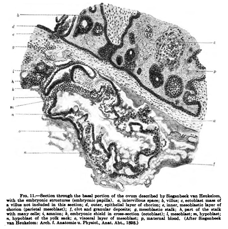
It was obtained from a woman who received some bums during an epileptic attack and died six hours later. An autopsy was performed fourteen hours after death. With the exception of the bums the body showed no noteworthy departures from the normal. The entire uterus was placed in 3 per cent, formalin for some days and was then washed and fixed in alcohol. Since the ovum was collapsed as the result of a slight tear, accurate measurements could not be made; but Van Heukelom estimates its meridian at about 16i mm., which would give a diameter of 5.1 mm. The ovum was completely covered with villi; the insertion of thirtyone of these could be counted in a meridional section, fifteen occurring on the basal portion of the section (the portion nearest the uterine wall), twelve at the opposite pole, and two on each side at the equator. The villi of the embryonic pole were better developed than those of the opposite pole, and those at the -equator were " especially heavy and thick." They varied greatly in length, some being small and short, the majority 0.75 mm. long and those at the equator as much as 1 mm. Some were little branched, others, especially the basal and equatorial ones, had many offsets, and some divided into numerous branches. The epithelial covering of the villi was composed of two layers; the outer showed no cell limits, so that we have to deal again with the Langhans layer and the syncytium, a cytotrophoblast and a spongiotrophoblast. All the large branches of the villi were connected peripherally by means of epithelial trabeculaB (ectoblast trabeculflB, cell columns) to form an epithelial shell (ectoblast or trophoblast shell) provided with large and small spaces. This shell was often very thin, consisting of only a single cell layer; at other times it was very thick and in its thicker parts there were peculiar blood lacuna, resting directly upon the maternal compacta. The remaining conditions resembled in essence those of the Von Herff ovum of Spec. At the basal wall of the ovum was the embryonic papilla (the embryonic structures) united to the chorion at a very acute angle by a stalk. A distinct allantoic duct was present and the amniotic cavity was lower than in the Von Herff ovum described by Spee. Only one of the sections through the embryonic shield is figured, and this is reproduced here as Fig. 11. In it, in the region of the primitive streak, a mass of cells is seen projecting beyond the surface of the shield; Van Heukelom suggested that this might be Hensen's node, a suggestion which I, as well as the author, would mark with a large note of interrogation. Nothing is said of an amniotic duct.
Somewhat older again is the ovum that I[16] described in 1890; it contained an embryonic shield with a well-developed primitive streak and was the first of this stage to be described. The ovum was expelled in an abortion with the egg capsule, which was lenticular in shape and measured 12 X ^i X 7 mm. ; it had a scar measuring 2i mm. Like all human ova of this period it was easily separated from the capsule, and measured SiX^iX^ mm. It had two areas free from villi : a larger one, measuring 6i X ^f ^^n., at the pole opposite the embryonic anlage, that is, the pole toward the lumen of the uterus; and a smaller one at the embryonic pole, situated exeentrically in front of the point of attachment of the belly stalk and measuring 2 mm. in diameter. The tissue of the Reichert scar showed neither glands nor blood-vessels, nor was it bounded by epithelium. The structure of the chorion and villi was the same as in the Spee ovum Von Herff, except that the syncytium was more strongly developed. The embryonic shield was about 1 mm. long and showed in the sections a well-developed primitive streak. The yolk sack liad a diameter of 1 mm. ajid showed numerous blood and bloodvessel aniagen. The endotlielial walls of the blood-vessels were already clearly distinguishable from the blood-corpuscles. There was an allantoic duct.
Fig. 11. Section through the basal portion of (ha ovum described by Siegenbeek van Heukelom.
The ova described by Merttens,[17] by Leopold in his Atlas, by Marchand and by Rossi need be mentioned here only in so far as they present especially striking peculiarities. Merttens had for study only four sections found aCoidentally in the investigation of a uterine curetting. Leopold in his ovum obtained by operation and estimated by him, " with doubtful accuracy," aCoording to Spee, to be seven to eight days old — it was undoubtedly older — found no distinct embryonic papilla, either on aCoount o£ unsatisfactory preservation, as Van Heukelom Euggesta, or, as Spee believes, because the ovuni was abnormal. At all events the ovum, whose diameter was 4 X 3-7 mm., need not be further considered here. Couceming the ova described by Marchand (1898) I may remark briefly that that author describes {Marburger 56., 1898, pp. 150-153), in an imperfectly preserved o\-um of the size of a pea, at that portion of the surface of the chorion where the remains of the embryonic anlage oCourred, a funnel-like depression of the surface of the chorion which led into a canal filled with syncytium; this he interpreted as the remains of an amniotic duct.[18]
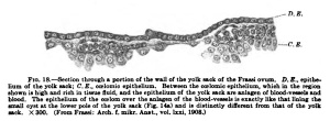
From two later publications by Marchand (" Beobachtungen an jungren menschlichen Eiem, Anat. Hefte, No. 67, vol. xxi, 1902, pp. 217-278; and " Einige '* Beobachtungen an jungen menschlichen Eiem," Verh. Anat. Ges., 1902) it may be noted that he found in one ovum[19] that the intervillous space was not filled with maternal blood, not withstanding that the blood-vessels of the neighboring portions of the mucous membrane were esoessively engorged. The intervillous space seemed to be separated from the cavity of the egg chamber peripherally by strong ectoblastic proliferations; blood vessels opening into the cavity of the egg chamber could not be found. From this it would follow that the blood must normally enter the intervillous space only at a later period of development; a precocious entrance of the blood may, according to Marchand, lead to an abortion. Also, according to Rossi Doria,[20] who described an ovum of less than 9X8 mm. contained in a completely closed egg capsule, an actual circulation of the blood does not take place at the beginning of the second week of development, because at that time the maternal blood has not yet gained access to the intervillous space; he regards the so-called prickle processes on the surface of the syncytium as a deposit formed from degenerated blood-corpuscles and the " scar " of the egg capsule as formed by regressive changes of the summit of the reflexa. Nothing is stated concerning the embryonic anlage, and the entire ovum was apparently little favorable for the settlement of important questions. We may now consider an ovum obtained by operation and studied by Frassi[21] under my direction. The entire uterus, removed per vaginam, was at once placed in a warm 5 per cent, solution of formalin; it remained there forty-eight hours and was then washed for twelve hours and finally passed through alcohols of gradually increasing strength. Only then was it opened, cut into portions, and these imbedded in celloidin, in which condition it came into the hands of Frassi. The ovum, together with the portion of the uterus that contained it, was cut into serial sections. The ovum and embryonic structures were undoubtedly normal.
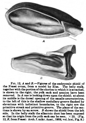
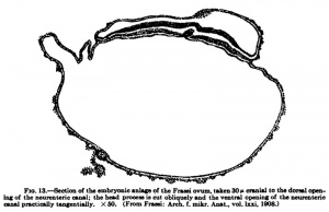
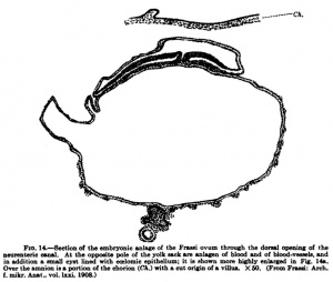
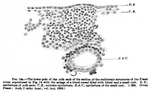
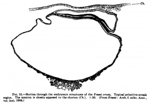
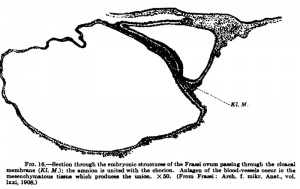
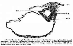
The diameter of the egg capsule parallel to the surface of the uterine lumen, in the plane of the sections, was 13 mm., perpendicular to this surface it was 5 mm. at the middle of the ovum; the corresponding diameters of the cavity of the ovum were 9.4 and 3.2 mm. A scar could not be detected in the decidua capsularis. The ovum was completely covered with villi, which were especially developed in the equatorial zone; their length varied between 0.5 and 1.9 mm. Both blood-vessels and glands opened into the intervillous space, but with regard to the latter it could be perceived that they had been laterally eroded, so that their communication with the space was secondary. It is remarkable that practically no blood was contained in the intervillous space, notwithstanding that bloodvessels opened into it; it must be that the blood had completely escaped during the operation. The Langhans layer, syncytium, and cell columns were present; and of these the Langhans layer and the cell columns may be regarded as cytotrophoblast and the syncytium as spongiotropboblast. The embryonic shield was cut somewhat obliquely; it showed the anlage of a well-developed primitive streak, at the anterior end of which was a neurenteric canal and at the posterior end the cloacal membrane. The section shown in Fig. 14 passed directly through the neurenteric canal. In front of the primitive streak is a flat medullary groove, bounded by still indistinct medullary folds. Anlagen of blood and blood-vessels occurred on the yolk sack. Anlagen of blood-vessels could be seen with certainty in the mesoderm of the chorion only in the nei^borhood of the insertion of the belly stalk; none could be detected in the mesodermal axes of the villi. Models were made of the embryonic structures as well as of the embryonic shield, but only those of the shield need be figured here, together with some of the sections.
The measurements, made on the model, were:
- Length of the embryonic shield 1.17 mm.
- Breadth of the embryonic shield 0.6 mm.
- Length of the primitive streak 0.5 mm.
- Diameter of the yolk sack,
- a. Greatest 1.9 mm.
- b. Least 0.9 mm.
The embryonic structures were attached to the inner surface of the chorion by a typical belly stalk, in which vessels could be made out.
We come now to the ovum Gle (Glaevecke), the careful study of which by Graf Spee has done so much to advance our knowledge of human embryology. It was an aborted ovum that was expelled, together with the entire uterine mucous membrane, five weeks after the cessation of the menses. The diameter of the egg capsule parallel to the surface of the decidua was 10 X 11 mm., and perpendicular to this, the thickness of the decidua basalis being included, 7.2 mm. The ovum was everywhere, but not very thickly, covered with villi. It was somewhat oval, its diameters being 8.5 X 10 X 6.5 mm. ; the last diameter is that perpendicular to the decidua basalis. The villi were covered by a Langhans layer (cytoltrophoblast) and a syncytium layer (spongiotrophobiast), and the cavity of the ovum had a horizontal diameter of 7.5 X 8 mm.
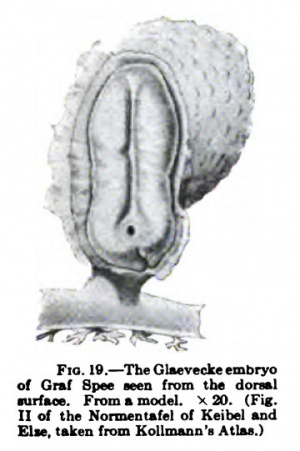
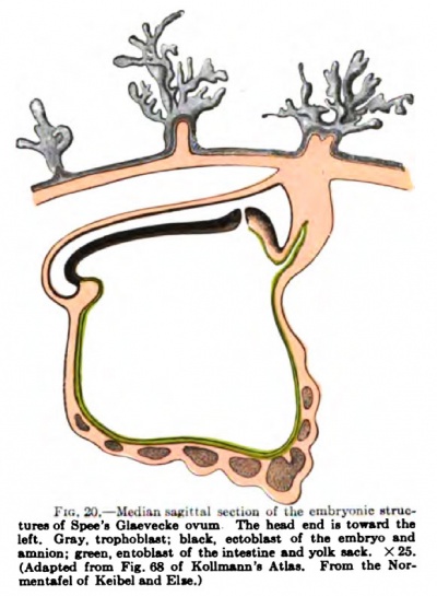
Fig. 19 shows the embryonic structure after a model by Graf Spee (from Kollmans "Atlas"), and Fig. 20 a median sagittal section of them. The amnion is represented as opened in Fig. 19. The primitive streak, which occupied halt of the germinal disk in the stage of development last described, is now limited to its posterior end, and thb is bent strongly downward. At the anterior end of the streak was a well-developed canatis neurentericus, and in front of this the medullary groove bounded by well-formed medullary folds.
The greatest direct length of the embryonic structures, measured from the anterior curve of the amnion to the attachment to the chorion before the embryo had been placed in alcohol, was 2 mm., and that of the germinal disk from the anterior curve of the amnion to the hind end of the primitive streak was 1.54 mm. Throughout this length the disk rested like a lid upon the yolk sack. Tbe average diameters of the germinal disk (i.e., the distance in a direct line between the lines of reflection of the germinal layers into the amnion and yolk sack) were: anteriorly, 0.704-0.741 mm.; at the middle and posteriorly, 0.665 mm.; in the region of the canalis neurentericus and primitive streak, 0.589 mm. ; and in the region of the twliy stalk, about 0.4 mm. The medullary plate, disregarding its curvature, presented its greatest diameter of 0.5170.57 mm. anteriorly; at its narrowest portion, about the middle of the germinal di^, it was 0.494 to 0,38 mm. in breadth. The hei^t of the belly stalk together with the amnion was 0.722 mm., the width of tbe ectoblast plate exclusive of the amnion was 0.361 mm., and that of the subjacent mesoderm mass was 0.209 mm. The lumen of the neurenteric canal was 0.024 mm. in diameter and that of tbe circular swelling seen surrounding it on surface view was 0.13 mm.
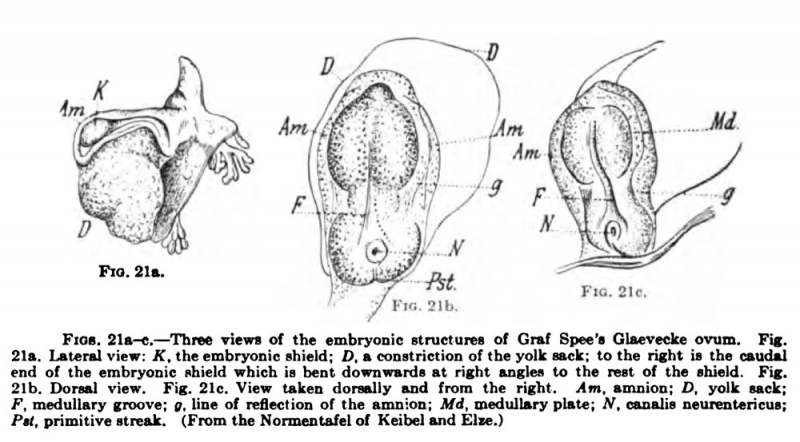
Of this important embryo some figures are here reproduced, some of tbe yet intact embryo as published by Spec in his paper of 1889 and some of sections of it. Fig 21a shows the embryonic stnicture seen from tbe side as an opaque object: K, the embryonic shield (germinal disk); D, a constriction of the yolk sack; to the left one sees the caudal end of the embryonic shield bent down at right angles to the rest of the shield. Fig. 21b shows the dorsal surface of the embryo, and in Fig. 21c it is diowii from the right and dorsally; both these drawings were made under direct illumination, the amnion being intaet bnt cleared with turpentine, while the germinal disk still remained opaque. The reflection of the amnion into the embryonic shield is indicated by g; at the hiuder end of the shield is the primitive streak (Psl) and in front of this is the opening of the neurenteric canal. In Fig. 21c the portion of the streak that is bent venlrally is also visible.
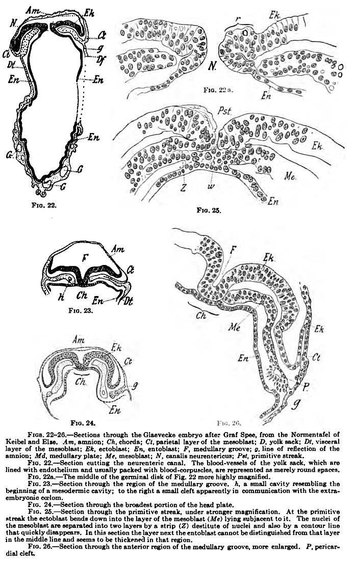
Figs. 22 and 22a represent a section passing tfarough the neurenteric canal ; the amnion, the yolk sack, and the relations of the germinal layers are shown. The distal portion of the yolk sack shows blood-vessels in course of development; the round spaces shown in that r^on represent vascular canals lined with endothelium and, for the most part, completely filled with young blood-corpuscles, not shown in the figure. The diameters of the vessels increase toward the distal surface of the yolk sack; on the right side the yolk sack was torn. Fig. 22& shows the middle portion of the section represented in Fig. 22 more strongly magnified.
Fig. 21. Three Views of the Glaevecke Embryo of Graf Spee
Fig. 23 is of a section through the region of the medullary groove. The chorda is included in the entoblast. The mesoblast shows to Ihe left a small cavity (h) resembling the cavity of a primitive segment in process of formation, and to the right is a narrow cleft in communication with the estra:-«mbryonic crelom.
Fig. 24 shows a section through the broadest portion of the head plate; it shows already a tendency toward the closure of the intestine. The primitive streak and primitive groove are represented more highly enlarged in Fig. 25. At the primitive streak the ectoblast bends down to unite with the layer of mesoblast subjacent to it. The nuclei of Ihe mesoblast are separated into two layers by a strip (Z) destitute of nuclei and also by a contour line that quickly disappears. The single layer that lies next to the entoblast cannot be distinguished from that layer in the middle of Ihe section, and in this region the entoblast appears thickened. P'ig. 26 shows Ihe half of a section passing through the medullary groove, greatly enlarged; at P it^shows the appearance of the pericardial cleft.
The embryo described by Elemod, a model of which has been reproduced by Friedr. Ziegler, is very similar to the Glaevecke embryo of Spec, but was not as well preser\-ed. It was obtained from a woman who had cohabited only during the night of November 6-7. The menslnaation expected on November 22 was omitted, the abortion occurred November 28.
Fig. 22-26.
In the fresh condition the entire ovum with its villi measured 10.0 X 8.2 X 6.0 mm. The villi had a length of 1.2-2.0 mm., their diameter being 0.3-0.8 mm. The embryonic shield was biscuit-shaped and measured in length from head cap of the amnion to the projecting caudal end 1.3 mm.; its breadth was 0.23 mm. in front and 0.18 mm. posteriorly. The closed amnion was continued into an amniotic duct. Further it may be remarked that Etemod describes the remains of a chorda canal in both the caudal and cranial ends of the chorda anlage, which is flattened and contained within the entoderm. Etemod's statements concerning the heart and the blood-vessels will be considered in the chapter dealing with those structures.
Since Spec's Glaevecke embryo showed indications of the commencing formation of the embryonic coelom and the primitive segments, it represents the final stage of the period of development we are here considering.
- ↑ G. Leopold : Ueber ein sehr junges menschliches Ei, Arb. Kgl. Frauenklinik, Dresden, vol. iv, Leipzig, 1906.
- ↑ Reichert : Beschreibung einer f riihzeitigen menschlichen Frucht im blaschenformijsren Bildungszustande, etc., Abh. Kgl. Akad. d. Wiss., Berlin, 1873.
- ↑ Thomas Wharton Jones : On the First Changes in the Ova of the Mammifera in Consequence of Impregnation and on the Mode of Origin of the Chorion, Philosoph. Transact. Royal Soc. London, 1837, p. 2.
- ↑ K. Breuss : Ueber ein menschliches Ei aus der zweiten Woche der Graviditat, Wiener med. Wochenschrift, 1877, pp. 502-504.
- ↑ Allen Thomson: Edinburgh Med. and Surg. Journal, vol. ii, 1839; and Froriep's Neue Notizen, vol. xiii, 1840.
- ↑ A. Kolliker: Entwicklungsgeschichte des Menschen, 1879.
- ↑ Franz Keibel and Curt Elze : Normentafel zur Entwicklungsgeschichte des Menschen, Jena, 1908.
- ↑ W. His: Anatomie menschlicher Embryonen, Leipzig, 1882, part ii, pp. 32 and 87 et seq. See also Mall, Joum. of Morph., vol. xix, p. 151.
- ↑ Bryce TH. and Teacher JH. Contributions To The Study Of The Early Development And Imbedding Of The Human Ovum 1. An Early Ovum Imbedded In The Decidua. (1908) James Maclehose and Sons. Glasgow.
- ↑ Hubert Peters: Ueber die Einbettung des menschlichen Eies und das friiheste bisher bekannte menschlichen Placentationsstadium, Leipzig und Wien, 1899.
- ↑ Those portions of the trophoblast which come into relation with the tissues of the uterine wall and take an active part in the implantation of the ovum and in the excavation of the egg chamber have been termed trophoderm by Minot (Charles S. Minot: The Implantation of the Human Ovum in the Uterus, Trans. Americ. Gynaecol. Soc., 1904).
- ↑ Carlo Giacomini: Un novo humano di 11 giorni, Giomale della Reale Academia di Medicina di Torino, vol. iii, anno 60, Fasc. 10-11, Torino, 1897.
- ↑ F. Marchand: Beobachtungen an jungen menschlichen Eiem, Anat. Hefte, vol. xxi, 1903.
- ↑ Mall FP. A contribution to the study of the pathology of early human embryos, (1900) Johns Hopkins Hosp. Rep., 9: 1-68. Also Mall FP. Normal plates of the development of vertebrates. Anat. Rec, (1908).
- ↑ Siegenbeek van Heukelom : Ueber die menschliche Plazentation, Arch, f . Anat. u. Physiol., Anat. Abt., 1898.
- ↑ Franz Keibel : Ein sehr junges menschliches Ei, Arch, f . Anat u. Physiol., Anat. Abt., 1890.
- ↑ J. Merttens: Beitrage zur normalen und patholog. Anat. der menschl. Placenta, Zeitschr. f. Geburtsh. u. G>-nakol., vol. xxx, 1894, and vol. ixsi, 1895. "0. I^opold: Uterus und Kind, Leipzig, 1897.
- ↑ In a later publication (Anat. Hefte, 1903, p. 223) he says: "We have to do therefore with a narrow canal traversing the choritn, which from Its opening at the surface is lined or, more properly, filled by a prolongation of the so-called surface epithelium, and ends blindly a short distance below the inner surface, just where the remains of the embryonic anlage occur." He ascribes the same significance to a depression on the surface of the chorion of an ovum of 14 X 3 mm diameters, which was laterally compressed by a blood-clot.
- ↑ "The ovum was obtained from the body of a woman who had died as the result of a gunshot wound. It was studied in situ, but the ovum and egg capsule were folded; the latter, according to Marchand, had a length of about 1.5 cm, and a breadth and depth of about 5-6 mm. The entire ovum was covered with branched villi. The embryo was completely disintegrated
- ↑ Tullio Jiossi Doria : Ueber die Einbettung des menschliehen Eies, studiert an einem kleinen Ei der zweiten Woche, Arch. f. Gynakol., 1905, vol. Issvi, pp. 433-505.
- ↑ L. Frassi : Ueber ein junges menschliches Ei in situ, Arch, f . mikr. Anat., vol. Ixx, 1907; and Weitere Ergebnisse des Studiums eines jungen menschlichen Eies in situ, ibid., vol. Ixxi, 1908.
| Historic Disclaimer - information about historic embryology pages |
|---|
| Pages where the terms "Historic" (textbooks, papers, people, recommendations) appear on this site, and sections within pages where this disclaimer appears, indicate that the content and scientific understanding are specific to the time of publication. This means that while some scientific descriptions are still accurate, the terminology and interpretation of the developmental mechanisms reflect the understanding at the time of original publication and those of the preceding periods, these terms, interpretations and recommendations may not reflect our current scientific understanding. (More? Embryology History | Historic Embryology Papers) |
Glossary Links
- Glossary: A | B | C | D | E | F | G | H | I | J | K | L | M | N | O | P | Q | R | S | T | U | V | W | X | Y | Z | Numbers | Symbols | Term Link
Cite this page: Hill, M.A. (2024, May 1) Embryology Book - Manual of Human Embryology 4. Retrieved from https://embryology.med.unsw.edu.au/embryology/index.php/Book_-_Manual_of_Human_Embryology_4
- © Dr Mark Hill 2024, UNSW Embryology ISBN: 978 0 7334 2609 4 - UNSW CRICOS Provider Code No. 00098G

