Book - Manual of Human Embryology 3
| Embryology - 1 Mar 2026 |
|---|
| Google Translate - select your language from the list shown below (this will open a new external page) |
|
العربية | català | 中文 | 中國傳統的 | français | Deutsche | עִברִית | हिंदी | bahasa Indonesia | italiano | 日本語 | 한국어 | မြန်မာ | Pilipino | Polskie | português | ਪੰਜਾਬੀ ਦੇ | Română | русский | Español | Swahili | Svensk | ไทย | Türkçe | اردو | ייִדיש | Tiếng Việt These external translations are automated and may not be accurate. (More? About Translations) |
Keibel F. and Mall FP. Manual of Human Embryology I. (1910) J. B. Lippincott Company, Philadelphia.
| Historic Disclaimer - information about historic embryology pages |
|---|
| Pages where the terms "Historic" (textbooks, papers, people, recommendations) appear on this site, and sections within pages where this disclaimer appears, indicate that the content and scientific understanding are specific to the time of publication. This means that while some scientific descriptions are still accurate, the terminology and interpretation of the developmental mechanisms reflect the understanding at the time of original publication and those of the preceding periods, these terms, interpretations and recommendations may not reflect our current scientific understanding. (More? Embryology History | Historic Embryology Papers) |
Keibel F. III. Segmentation in Keibel F. and Mall FP. Manual of Human Embryology I. (1910) J. B. Lippincott Company, Philadelphia.
III. Segmentation
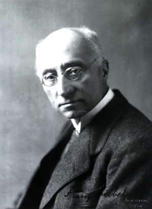
By Franz Keibel, Freiburg i. Br.
The segmentation stages of the human ovum have not yet been observed. We may with certainty assume that the early stages of fertilization are passed through during the passage of the ovum through the tube, but whether the entire segmentation takes place during this passage, or in what stage of segmentation the ovum reaches the uterus, cannot even be conjectured. In mammals there are apparently differences in this respect. The time, also, that the human ovima requires for the passage of the tube is very difficult to estimate; according to the data obtained from other manunals it cannot well be believed that the uterus is reached before the fifth day. Similarly, it is unknown whether the ovum becomes imbedded in the mucous membrane immediately after it has reached the uterus. That it is possible by good fortune and persistency yet to observe segmenting human ova in the tube is shown by the observations of Letheby [1] and Hyrtl (in a work by Bischoff and in Froriep's "Neue Not.," 1852, No. 603), who discovered ova in the tube, althouja:h they were not able to make observations of the segmentation, partly on account of the imperfections of their technic and partly because the ova were unfertilized. The relative certainty with which experienced embryologists are able to-day to obtain the segmentation stages of even large mammals is an encouragement for further efforts in this direction. The force which propels the ovum through the tube into the uterus is the ciliary action of the tubal epithelium, and injury to this epithelium mav be the cause of the retention of the ovum in the tube or in a diverticulum of it and so the cause of a tubal pregnancy. If the ciliary current is not impaired, the ova are readily driven over any diverticula that may exist (Kromer[2]).
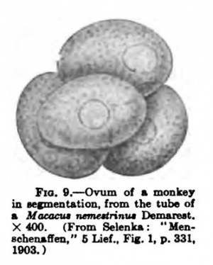
It may be regarded as quite eertaia not only that the human ovum undergoes a segmentation quite similar to that of the other mammals, but also that it is a secondary total segmentation. The ancestors of the human species, like those of other mammals, must have once possessed yolk-laden meroblastic ova. A separation of the segmentation cells ccording to their developmental potencies has been variously postulated for the earlier stages of the mammalian segmentation, but conclusive evidence for this is lacking, the observations hitherto made not being capable of such an interpretation. This is true also of the observations which have been supposed to indicate the existence of a gastrulation process in the later stages, but this question will be considered in the chapter dealing with the formation of the germ layers and the gastrulation problem. Finally, it may be noted here that Hubrecht has recently succeeded in finding the ovum of a monkey in segmentation. It has been figured in ia KsmeDtatioD. from the tuba of the posthumous paper by Selenka which I have X wo (" MenschenafEen," 5 Lief., Zur vergleich ended Keimesgeschichte der Primaten, Wiesbaden, 1903) and is reproduced here (Fig. 9), since it is the only primate ovum in a segmentation stage at present known. Selenka states concerning this ovum, which was found in serial sections of a tube of a Macaacus nemestrinus from Java: "At about the middle of the oviduct was the ovum, having a diameter of 0.04 mm. and loosely attached to the somewhat frayed out ciliated cells. The largest of the approximately ripe ovarial ova were of about the same size. Four segmentation cells of about equal size are clearly to be distinguished; two of these (the central and left upper ones in the figure) are irregularly oval, the other two are almost spherical. The cells are naked; no trace of an enclosing membrane is to be observed. The shrinkage which tie tissues of the oviduct show suggests the idea that the segmenting ovum no longer retains its natural condition. It is, however, of importance to note what the preparation reveals: The segmentation begins in a manner rimilar to that of other higher mammals, and it is probable that it is completed as soon as the ovum has entered the uterine enlargement" This last conclusion I cannot accept.
- ↑ H. Letheby: An Account of Two Cases in which Ovules or Their Remains Were Discovered in the Fallopian Tubes of Unimpregnated Women who Had Died during the Period of Menstruation, Philos. Transact. Royal Soc. London, 1852 (altogether unsatisfactory). See also Jroriep's Neue Notizen, 1852, No. 603. ' Th. L. W. Bischoff : Beitrage zur Lehre von der Menstruation und Bef nichtung, Zeitschrift fiir rationelle Medizin, neue Folge, vol. iv, 1854.
- ↑ P. Kromer: Untersuchungen iiber den Bau der menschlichen Tube, Leipzig, 1906.
| Historic Disclaimer - information about historic embryology pages |
|---|
| Pages where the terms "Historic" (textbooks, papers, people, recommendations) appear on this site, and sections within pages where this disclaimer appears, indicate that the content and scientific understanding are specific to the time of publication. This means that while some scientific descriptions are still accurate, the terminology and interpretation of the developmental mechanisms reflect the understanding at the time of original publication and those of the preceding periods, these terms, interpretations and recommendations may not reflect our current scientific understanding. (More? Embryology History | Historic Embryology Papers) |
Glossary Links
- Glossary: A | B | C | D | E | F | G | H | I | J | K | L | M | N | O | P | Q | R | S | T | U | V | W | X | Y | Z | Numbers | Symbols | Term Link
Cite this page: Hill, M.A. (2026, March 1) Embryology Book - Manual of Human Embryology 3. Retrieved from https://embryology.med.unsw.edu.au/embryology/index.php/Book_-_Manual_of_Human_Embryology_3
- © Dr Mark Hill 2026, UNSW Embryology ISBN: 978 0 7334 2609 4 - UNSW CRICOS Provider Code No. 00098G


