Book - Manual of Human Embryology 19-4
| Embryology - 27 Apr 2024 |
|---|
| Google Translate - select your language from the list shown below (this will open a new external page) |
|
العربية | català | 中文 | 中國傳統的 | français | Deutsche | עִברִית | हिंदी | bahasa Indonesia | italiano | 日本語 | 한국어 | မြန်မာ | Pilipino | Polskie | português | ਪੰਜਾਬੀ ਦੇ | Română | русский | Español | Swahili | Svensk | ไทย | Türkçe | اردو | ייִדיש | Tiếng Việt These external translations are automated and may not be accurate. (More? About Translations) |
Felix W. The development of the urinogenital organs. In Keibel F. and Mall FP. Manual of Human Embryology II. (1912) J. B. Lippincott Company, Philadelphia. pp 752-979.
| Historic Disclaimer - information about historic embryology pages |
|---|
| Pages where the terms "Historic" (textbooks, papers, people, recommendations) appear on this site, and sections within pages where this disclaimer appears, indicate that the content and scientific understanding are specific to the time of publication. This means that while some scientific descriptions are still accurate, the terminology and interpretation of the developmental mechanisms reflect the understanding at the time of original publication and those of the preceding periods, these terms, interpretations and recommendations may not reflect our current scientific understanding. (More? Embryology History | Historic Embryology Papers) |
Cite this page: Hill, M.A. (2024, April 27) Embryology Book - Manual of Human Embryology 19-4. Retrieved from https://embryology.med.unsw.edu.au/embryology/index.php/Book_-_Manual_of_Human_Embryology_19-4
- © Dr Mark Hill 2024, UNSW Embryology ISBN: 978 0 7334 2609 4 - UNSW CRICOS Provider Code No. 00098G
IV. Development of the External Genitalia
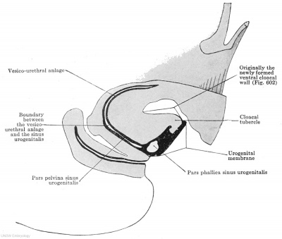
The parent tissue for the external genitalia is the cloacal tubercle. It has already been pointed out (p. 874) that this appears between the umbilicus, the coccygeal tubercle and the two lower limbs in an embryo of 13 mm (Figs. 637, 638). We may distinguish on it an oral and an anal, a right and a left slope. Along the anal slope, from the summit to the base, there is, in the interior of the tubercle, the pars phallica of the urogenital sinus (Fig. 638), whose ventral wall, formed by the urogenital membrane, abuts upon the surface of the cloacal tubercle (Fig. 638). The tubercle is originally a paired structure, lying on either side of the cloacal membrane (Fig. 601), but the paired condition is converted into an unpaired one by the elevation of the entire anterior abdominal wall (Figs. 601, 637).
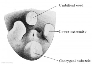
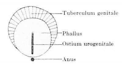
Upon this cloacal tubercle the sexual organ, the phallus, is seated like a tower on a hill. The base of the phallus thereby comes to lie excentrically on the cloacal tubercle, occupying the whole of its anal slope and a part of the right and left ones (Fig. 639). The tubercle is thus divided into the almost circular base of the phallus and the semilunar genital tubercle (Fig. 639). This latter represents an originally unpaired structure, lying cranial to the phallus and surrounding it on two sides ; from it there are formed later the two genital swellings. The genital tubercle is separated from the phallus in female embryos by a deep groove which is the anlage of the later sulcus nympholabialis, the groove between the labia majora and minora. In male embryos this groove is absent from the beginning.
Fig. 638. Sagittal section through the caudal end of the body of an embryo of 24 mm vertex-breech length. (Embryo Hal. 1, from the collection of the I Anatomical Institute, Vienna.) The cloacal tubercle projects between the umbilicus and the coccygeal tubercle. The pars phallica of the sinus urogenitalis runs along its anal slope.
Fig. 639. Diagrammatic representation of the division of the cloacal tubercle into the phallus and genital tubercle.
The Indifferent Phallus
The quickly growing phallus assumes the penial form at an early stage in both sexes, and at the beginning of the third month a transitory sexual difference occurs in the direction of the phallus, which in female embryos bends downwards, while in male embryos it retains its direction more or less perpendicular to the long axis of the body (Herzog 1904).
The pars phallica of the urogenital sinus, which is contained within the phallus, behaves differently in different parts, that part which corresponds to the future glans becoming perfectly solid and forming the urethral plate, while the part corresponding to the rest of the penis remains hollow, and by destruction of the urogenital membrane breaks through to the exterior throughout its entire length; it forms the trough-like orificium urogenitale primitivum. As soon as the coronary sulcus of the glans is developed in embryos of 21 mm, it lies at about the boundary between the anterior circumference of the orificium urogenitale and the urethral plate.
The glans is marked off as a spherical protuberance in embryos of 26 mm.
The Sexual Differentiation
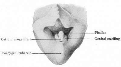
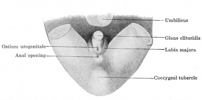
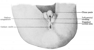
The beginning of the sexual differentiation can hardly be assigned to a definite period, since it takes place quite gradually and even in advanced stages presents difficulties to diagnosis. We base our diagnosis on the position of the ostium urogenitale relative to the coronary sulcus on the one hand and the anal opening on the other. Male embryos are distinguished by the distal ("distal" has reference here to the base of the phallus) circumference of the ostium urogenitale always lying in the coronary sulcus, while the proximal circumference becomes more and more distant from the anal opening. Female embryos may be recognized by the urogenital opening retreating away from the coronary sulcus by the gradual closing of its distal part, while its proximal part always lies close to the anal opening; the differentiating moment is, accordingly, both positive and negative in each sex. In Fig. 640 are shown the external genitalia of an embryo in which a determination of the sex from the external genitalia is impossible. The urogenital opening is on the anal slope of the phallus, whose projecting summit already shows the anlage of the glans. The distal circumference of the opening reaches the sulcus, the proximal one still lies close in front of the anal opening; the first fact is positive for male embryos, the second positive for female ones, and consequently these genitalia are to be termed indifferent. Fig. 641 shows the female differentiation: the phallus has become the clitoris; the urogenital opening lies close in front of the anal opening, and from its distal circumference a shallow groove, the urethral groove, extends to the coronary sulcus. A wandering away from the sulcus coronarius and a persistence of the relations to the anal opening are both female characters. Fig. 642 shows the male type. The urogenital opening is preserved throughout its entire length, its distal circumference reaches the sulcus coronarius, the proximal one has separated from the anal opening: a wandering away from the anal opening and the preservation of the relation to the coronary sulcus are both male characters.
Fig. 640. The indifferent external genitalia of an embryo of 28 mm greatest length. (From a photograph kindly furnished by Professor R. Meyer, Berlin.) The cloacal tubercle is divided into the phallus and the genital tubercles or swellings. The ostium urogenitale and anus are still close together.
Fig. 641. Female external genitalia of an embryo of 32.5 mm greatest length. (From a photograph kindly furnished by Professor R. Meyer, Berlin.) The phallus has become the clitoris, the glans and coronary sulcus are recognizable. The ostium urogenitale has preserved its relation to the anal opening, but no longer reaches the sulcus coronarius glandis.
Fig. 642. Male external genitalia of an embryo of 3.5 months. (From a photograph kindly furnished by Professor R. Meyer, Berlin.) The phallus has become the glans penis, the scrotal swellings are fully developed and the unpaired scrotal area is indicated. The ostium urogenitale and ostium anale have separated, the ostium urogenitale extends to the sulcus coronarius glandis.
Development of the Penis and Scrotum
The most striking characteristic of the transformation of the indifferent phallus, into the penis is the great elongation of the male organ. The formation of the penis takes place in such a way that the glans and the distal portions of the shaft of the penis are first formed. It has been pointed out that the sulcus coronarius glandis appears in the indifferent stage in embryos of 21 mm. and that embryos of 26 mm already possess a distinctly projecting glans. After the glans and distal portions of the shaft are formed, the formation of the rest of the shaft occurs, and these newlyformed portions gradually raise up the already formed glans and distal portions of the shaft. The glans is passive to the extent that it only enlarges in proportion to the entire growth. Along with the glans and the distal portions of the shaft the coronary sulcus and the ostium urogenitale are also raised up. The form, size and position of the latter remains the same from the beginning to the end of the growth of the penis and is in all stages of development a distally broadened slit (Fig. 642), which extends to the sulcus coronarius glandis.
The urogenital sinus, accordingly, appears at once in its whole length; it does not grow by the gradual closure of a groove along the anal wall of the penis until it reaches the glans, but its growth in length keeps pace with that of the penis. The ostium urogenitale, accordingly, already represents the primitive external orifice of the sinus urogenitalis.
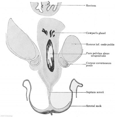
Since in this growth the first-formed parts of the penis are pushed out passively, we must assume the existence of a basal zone of growth lying to the pelvic side of the urogenital opening, that is to say, in the neighborhood of the pars pelvina of the urogenital sinus. The pars pelvina is, accordingly, with reference to the growth, the active portion of the urogenital sinus, and the pars phallica the passive portion. The basal growth in the pars pelvina must produce a new area, interposed between the base of the penis and the anal opening, and the best name for this is the unpaired scrotal area (Fig. 642). In embryos of 60 mm. head-foot length this becomes raised up in toto and forms the unpaired scrotal swelling, into which the two genital swellings, which we now must term the scrotal swellings, extend from above. Since the middle of the area is connected by dense connective tissue with the connective tissue of the pars pelvina, the projecting scrotal swellings gradually draw out this connective tissue to a band and so the septum scroti is formed, it being at all times only a connective tissue structure. To its stretching it offers a certain amount of resistance, and consequently, there is formed at the surface of the scrotal area a median depression (Fig. 643), which, from the beginning, is the only external division of the unpaired scrotal swelling into a right and left half, while the septum scroti effects a complete internal division. As soon as the descensus is complete this unpaired scrotal area alone forms the scrotal sack, the paired scrotal swellings to the right and left of the penis fading out into the surrounding areas.
Fig. 643. Transverse section of a male embryo of 70 mm head-foot length (embryo R. Meyer 267, collection of Professor R. Meyer, Berlin; slide 67, row 1, section 3), below the symphysis pubis. X ca. 23. From behind forwards the following parts are cut: rectum, pars pelvina of the urogenital sinus and scrotum. From the pars pelvina Cowper's glands have developed and bet ween A the mesenchyme of the urogenital sinus and the epithelium of the scrotum there is stretched the septum scroti.
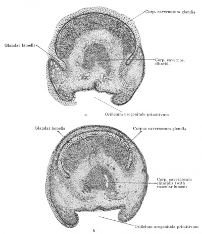
The primitive glans becomes divided into two portions (Fig. 644 a and b) by an epithelial cylinder that grows in from the epithelium of the external surface; the two parts are an outer cylindrical mantle, the praeputium, and an inner sphere, the glans in the narrower sense. The glandar lamella (Fleischmann 1907), as we shall term this epithelial cylinder, is not a closed tube, but is imperfect on the anal surface ; by this opening the prasputium and the glans in the narrower sense remain in connection, the frenulum prceputii is a persistent portion of the mesenchyme of the primitive glans. The solid glandar lamella becomes hollowed out by the degeneration of its central cells, the cavity breaks through to the exterior and so the space between the prepuce and the glans in the narrower sense is formed. The glandar lamella makes its appearance in embryos of 50 mm. head-foot length.
Fig. 644 a and b. Two sections through the glans clitoridis of a human embryo of 80 mm head-foot length. (Embryo R. Meyer 266, from the collection of Professor R. Meyer, Berlin; slide SS, row 2, section 5, and row 2, section 3.) From the surface epithelium a solid lamella, the glandar lamella, grows into the mesenchyme of the glans and separates the glans proper from the preputium.
The corpora cavernosa appear as four portions, which, enumerated in the order of their appearance, are: the two corpora cavernosa penis, the corpus cavernosum glandis and the corpus cavemosum urethrae. They are formed from the young connective tissue by a dense aggregation of the mesenchyme cells, which are packed so closely together that the anlage has the appearance of being perfectly solid; only after some time greatly scattered vascular lumina occur and these later enlarge; between the first formed vessels new ones are formed and the characteristic structure of the spongy tissue is acquired.
The corpora cavernosa penis are united together at their apices and along their oral surfaces.
The first traces of a corpus cavernosum penis are to be found in embryos of 14.75 mm, the corpus cavernosum glandis occurs in embryos of 22 mm greatest length. A compact layer of mesenchyme cells occurs around the epithelial lining of the sinus urogenitalis, at an early period, but a definite delimitation of a corpus cavernosum urethra? is first seen in embryos of 70 mm head-foot length. The first vascular lumina occur in the glans in embryos of 28 mm greatest length and in the corpora cavernosa penis in embryos of 70 mm head-foot length.
Development of the Clitoris and of the Labia Majora and Minora
In the female there is formed from the phallus the clitoris, the frenulum clitoridis and the labia minora. Since the penis develops from the entire phallus it cannot be considered exactly homologous with the clitoris. The clitoris corresponds to the entire glans penis and the oral slope of the penis shaft, the f renula clitoridis and the labia minora correspond to the anal slope of the penis shaft (Fig. 648).
The phallus of the female shows at first the same amount of growth as that of males of the same age, indeed, it may even be longer. The urogenital opening always remains at the base of the phallus and must, therefore, descend anally along the anal surface of the clitoris; a groove, the urethral groove, between it and the sulcus coronarius of the glans shows the former extent of the urogenital opening. We may therefore recognize in the female also an ostium urogenitale primitivum and definitivum. The principal growth in length, which in the male occurs in the neighborhood of the pars pelvina of the urogenital sinus, is entirely wanting in the female and consequently after a short period of growth there is cessation of the growth in length of the clitoris and the urogenital opening remains in the neighborhood of the anal opening ; between the two there develops a short perineum, but never a scrotal area.
The two genital swellings on this account are not prolonged anally to form an unpaired scrotal tubercle and consequently they do not fade out as in the male, but persist as the labia majora. The middle part of the genital tubercle becomes the mons Veneris.
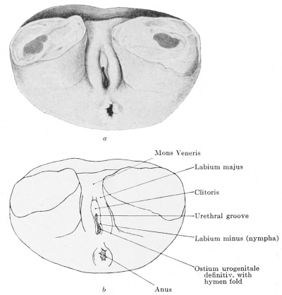
The labia minora or nymphae arise in their oral parts from the margins of the urethral groove, in their anal parts from the margins of the ostium urogenitale definitivum, that is to say, they arise from the entire anal surface of the phallus with the exception of the short portion that forms the anal surface of the clitoris ; they extend from the anal periphery of the ostium urogenitale definitivum to the sulcus coronarius glandis. The oral and anal portions do not arise simultaneously; the time of the first development of the oral portions cannot be given, for they are present as the swollen margins of the urethral groove so soon as the urogenital opening has broken through (Fig. 645) ; the swelling of the margins of the ostium urogenitale definitivum and consequently the anlage of the anal portions of the nymphae begins in embryos of 223 mm. vertexbreech length (measured over the nape and back) and 162 mm. in greatest head circumference; it remains, however, quite insignificant for a long time and it is only in embryos of 347 mm. vertexbreech length (measured over the nape and back) that one can speak of labia minora in the neighborhood of the ostium urogenitale definitivum, but even in this embryo the oral portions of the nymphae are still almost three times as high as the anal portions (Fig. 627). In embryos of 386 mm. vertex-breech length (measured over the nape and back) and 324 mm. in greatest head circumference the form of the adult nymphae is acquired and no difference obtains between the oral and anal portions.
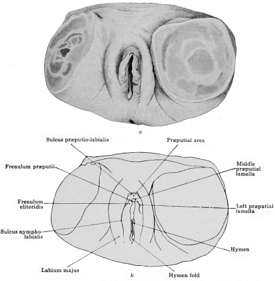
The formation of the glandar lamella proceeds somewhat more complicatedly in the female than in the male. Indeed, we must recognize in the female three glandar lamellae, an unpaired, ringshaped one that penetrates at the tip of the clitoris, and two paired lateral ones, that lie in the oral portions of the nymphae (Fig. 646). All three lamellae together form a narrow band, which encloses the glans clitoris and the later formed frenula clitoridis (Fig. 646). The middle ring-shaped lamella I found macroscopically for the first time in an embryo of 173 mm. vertex-breech length (measured over nape and back) and with a greatest head circumference of 163.5 mm. ; it corresponds to the male glandar lamella, but is less complete than this, forming only somewhat more than the half of a cylindrical mantle (Fig. 644 a and b). The frenulum praeputii is consequently broader than in the male. The formation of the cavity in the glandar lamella takes place practically as in the male and by it the glans clitoridis and the middle part of the praeputium are separated. Whether the two lateral lamellae, like the middle one, are actually formed as lamellae and later form a cavity, or whether they occur as grooves from the beginning I cannot state positively. Both the lateral grooves appear simultaneously in embryos of 347 mm. vertex-breech length (measured over nape and back) ; they begin at the left or right periphery of the middle groove and run obliquely over the outer surfaces of the nymphae to the sulcus nympholabialis (Fig. 646). By them the oral portions of the nymphae are divided lengthwise and from now on we may recognize an undivided portion, the definitive symphse and a genital portion, whose lateral parts give rise to the lateral parts of the praeputium, while its medial part becomes the frenulum clitoridis. Both frenula may be followed orally to the clitoris ; they are incompletely separated from the frenulum praeputii by the ends of the circular glandar lamella (Fig. 646). Although actually separated, the three glandar lamellae give the impression of a single structure; between them and the sulcus prasputic-labialis (Fig. 646) lies the praeputial area (Fig. 646), which is composed of the greater part of the anal and the right and left surfaces of the phallus. This preputial area Incomes raised up like a wall along the boundary formed by the three glandar lamellae, and so forms a rather deep groove (sulcus praeputio-clitoridis) between itself and the glans and frenula clitoridis. When the wall has reached a certain height it advances over the sulcus prEeputio-clitoridis, the clitoris and its frenula. The extent of this advancement varies individually; when it is slight the clitoris remains free, when it is greater the clitoris may be completely covered. What is termed the praepntrum in systematic anatomy is formed from this fold of the preputial area and consequently the prnaputium of the adult is a single structure, if, however, it is drawn backwards one may see the sulcus praeputio-clitoridis and at the bottom of this the three grooves formed by the splitting of the glandar lamellae. The adult praeputium is therefore a pseudopraBputium, the true praeputium lies at the bottom of the sulcus praaputio-clitoridis ; we may preferably speak of a clitoris sack. From what has been said it follows that the praeputium penis cannot be directly homologized with the praeputium clitoridis ; the praeputium penis is equivalent only to the small, middle, ring-shaped, true praeputium clitoridis.
Fig. 645 a and b. External genitalia of a female embryo of 27.2 mm vertex-breech length and 21.3 mm in greatest head circumference. X 13^- The phallus is enclosed by two genital swellings (labia majora). Its shorter oral surface is lost in the mons Veneris, the longer anal surface fading out into the posterior commissure of the labia majora. The summit of the phallus is occupied by the slightly projecting glans; a sulcus coronarius glandia cannot be seen. Anal from the glans is the cleft-like ostium urogenitale, that leads in its oral two-thirds into a shallow groove, the urethral groove, and in its anal third into the sinus urogenitalis. Into this sinus the hymen fold projects, but it does not reach the margin of the ostium urogenitale.
Fig. 646 a and b. External genitalia of a female embryo of 34.7 cm vertex-breech length (measured over nape and back). XIJ^. On the oral surface of the phallus and in the oral portions of the two nymphcc three glandar lamella? are developed, a middle unpaired one and two lateral paired ones. The three lamellae bound an area oralwards, the praeputial area, whose oral limit forms a groove (sulcus praeputio-labialis i between itself and the labia majora; from the praeputial area the clitoris sack develops. Between the two nymphs the hymen fold projects; it has in this case the unusual spoon-like form.
The formation of the corpora cavernosa takes place in the following order: corpus cavernosum clitoridis, corpus cavernosum glandis ; a corpus cavernosum urethrae does not occur, since the growth in length of the pars pelvina of the urogenital sinus is omitted. The formation of the corpora takes place as in the male.
The corpus cavernosum clitoridis appears in embryos of 19.4 mm, the corpus cavernosum glandis in embryos of 28 mm. greatest length and probably earlier.
Homologies of the Male and Female External Genitalia
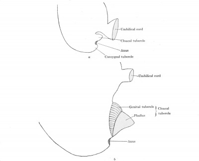
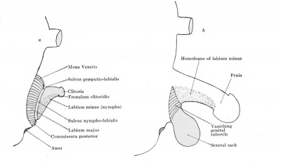
I shall now give a resume of the entire development of the external genitalia and shall endeavor to represent it in four diagrams. (1) The cloacal tubercle forms between the umbilicus and the oral periphery of the coccygeal tubercle (Fig. 647 a). (2) The phallus is placed excentrically on the cloacal tubercle, as one erects a tower on a hill. Thereby the cloacal tubercle is divided into the base of the phallus and the semilunar tuberculum genitale (Figs. 639 and 647 b). The indifferent condition lasts up to this period. (3) In the female the phallus becomes separated from the tuberculum genitale by a groove, sulcus praeputio-labialis and nympholabialis. The latter surrounds the phallus and so forms a circular swelling from which are formed: (1) the mons veneris from the oral portion, (2) and (3) the right and left labia majora from the right and left portions, and (4) the posterior commissura labiorum, from the anal portion (Fig. 648 a). On the phallus the glans becomes separated from the. shaft by a groove, the sulcus coronarius glandis. From this sulcus the urethral groove runs along the middle of the anal surface to the base of the phallus, where it is continued into the ostium urogenitale definitivum (Fig. 645). On both sides of the urethral groove and of the ostium urogenitale the anal surface of the phallus becomes enlarged and forms the labia minora (stippled in Fig. 648 a). The rest of the phallus becomes the clitoris and clitoris sack. (4) In the male the tuberculum genitale fades out in all its parts and in its place there is formed at the anal periphery of the phallus, from the unpaired scrotal area, a new swelling, the unpaired scrotal sack. The entire phallus becomes the penis, in which, as in the female, the glans becomes marked out by the sulcus coronarius glandis.
According to this account the clitoris corresponds only to the glans and the neighboring oral portions of the penis shaft (white in Fig. 648 b) ; a portion that corresponds to about the basal threequarters of the penis shaft is not formed in the case of the clitoris. The primitive ostium urogenitale of the male is not equivalent to that of the female, since in the male the ostium retains its place, while in the female it is displaced basally, or at least is shortened along the urethral groove. A homologue of the labia major a exists in the male only during development in the paired scrotal swellings. No structure corresponding to the unpaired scrotal swelling is formed in the female; we must take the commissura posterior to be its equivalent. The homologue of the labia minora of the female is not formed in the male, the anal surface of the penis between the sulcus coronarius glandis and the base is the corresponding region (stippled in Fig. 648 b).
Fig. 647 a and b.
Fig. 648 a and b.
Further Development of the Sinus Urogenitalis
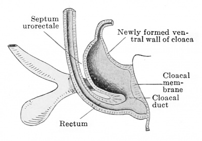
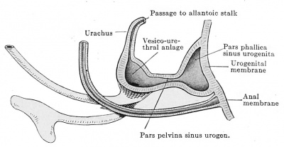
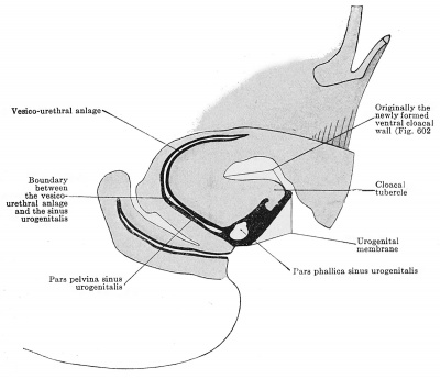
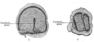
In the preceding sections of this chapter it has been impossible to avoid occasional allusions to the development of the urogenital sinus ; I shall now give an account of the entire development. The sinus is formed by a division of the cloaca into three parts in the frontal direction. The first division separates off the rectum (Figs. 649 and 650), the second divides the remains of the cloaca into the vesico-urethral anlage and the pars pelvina of the sinus urogenitalis on the one hand, and the pars phallica of the sinus urogenitalis on the other (Fig. 651). The sinus begins at Miiller's tubercle and ends at the tip of the phallus (Fig. 652). It is laid down at the beginning throughout its entire length and from the beginning is divided into its two portions, the pars pelvina and the pars phallica. The lumen of the pars phallica, which was formerly the lumen of the cloaca, and is present from the beginning, persists only in the basal portion of the cloaeal tubercle, vanishing in the distal portion by the right and left walls of the pars phallica being pressed into contact and finally fusing (embryos of from 24 to 29 mm. greatest length). The hollow portion of the pars phallica breaks through to the outside by the resorption of its ventral wall throughout its entire extent and so the ostium urogenital primitivum is formed. The solid part, which occurs only in the region of the glans, is termed the urethral plate. . From its mode of origin the urethral plate must extend to the summit of the cloacal tubercle and must be fused with the anal slope of the tubercle throughout its entire extent; the region of fusion corresponds to the cranial portion of the former cloacal membrane. Seen in a transverse section of the phallus the urethral plate extends from the anal periphery only to about the centre and, accordingly, it divides into two parts only the anal portion of the phallus (Fig. 653 a) ; at the summit the division is incomplete (Fig. 653 b), the urethral plate here standing in connection with the so-called epithelial horn, a meaningless epithelial growth (Fig. 652).
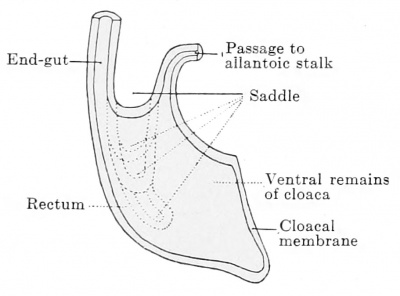
Fig. 649. The cloaca is divided into the rectum and the ventral remains of the cloaca by the gutterlike saddle sinking down between the end-gut and the allantois stalk.
Fig. 650. The division of the cloaca into the rectum and the ventral remains of the cloaca is almost completed, between the two there remains for a time a cloaeal duct, as an undivided portion of the cloaca (see also the explanation to Fig. 603).
Fig. 651. The ventral remains of the cloaca is drawn out in the ventro-dorsal direction and we maydistinguish in it four walls, — cranial, ventral, caudal, and dorsal. The cranial wall is depressed so that it approaches the caudal one. Thereby the ventral remains of the cloaca is incompletely divided into three parts, a dorsal broad part, the vesico-ure.hral anlage, a middle narrow part, the pars pelvina, and a ventral broad part, the pars phallica.
Fig. 652. The division of the ventral remains of the cloaca is completed. The vesico-urethral anlage and the pars pelvina of the sinus urogenitalis are narrowed in their dorso-ventral diameters, broadened in their frontal ones. The pars phallica remains broad in the sagittal diameter, but is narrowed in the frontal one, so that the lumen is abolished in the region of what will later be the glans; from the two walls an epithelial plate, the urethral^plate, is formed. The lumen is retained, corresponding to the extent of the ostium urogenitale (not cut in the section figured).
Fig. 653 a and b. Two sections through the clitoris of an embryo of 60 mm head-foot length. (Embryo R. Meyer 268, from the collection of Professor R. Meyer, Berlin; slide 66, row 2, section 1 and slide 67, row 1, section 2.) The urethral plate in section a extends from the anal surface of the clitoris only to the centre. In section 6, which passes through the tip, the clitoris is completely divided by the urethral plate.
In both sexes the ostium urogenitale primitivum is broadened out in a trough-like manner at the tip of the phallus (Fig. 642 and 654), and at the point where it breaks through there is a sharp distinction between the endodermal epithelium of the sinus and the ectodermal epithelium of the surface.
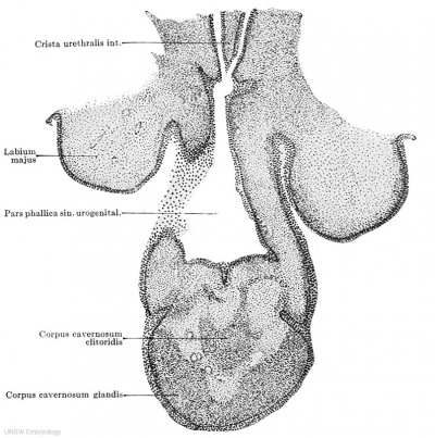
With the growth of the phallus and penis described above there is also, of course, growth of the sinus urogenitalis. In Fig. 654 one sees almost the entire length, perhaps it would be better to say shortness, of the sinus urogenitalis. It begins above with the crista urethralis, which runs downwards from Miiller's tubercle, and ends in the urethral plate. The pars pelvina and the pars phallica were originally of the same length, but during the growth in length of the entire region the pars phallica remains unchanged and the pars pelvina becomes enormously enlarged. The ostium urogenitale primitivum always extends to the sulcus coronarius glandis, its sagittal diameter remains always the same. The entire length of the sinus depends solely on the enlargement of the pars pelvina, which carries before it the pars phallica or the ostium urogenitale primitivum and the urethral plate. It has thus been shown that the urogenital sinus extends from Muller's tubercle to the primary ostium urogenitale and is entirely of entodermal origin.
Fig. 654. Longitudinal section of the clitoris of an embryo of 60 mm head-foot length. (Embryo H. Meyer 268, from the collection of Professor R. Professor R. Meyer; slide 64, row 2, section 1.) X 50. The section cuts the sinus urogenitalis lengthwise from the crista urethralis int. to the ostium urogenitale. The lumen of the sinus is enlarged at its end to a triangular basin, on whose distal wall is the urethral plate. To the right and left are the genital swellings, the anlagen of the labia majora, which are becoming separated from the clitoris.
The ostium urogenitale primitivum is the definitive one in neither of the sexes ; we know from macroscopic anatomy that the sinus of the male opens at the summit of the glans. This elongation is brought about by the closure of the ostium primitivum and the development of the glandar portion of the sinus from the urethral plate. I have not observed these processes myself and therefore follow the account given by Herzog (1904). In an embryo of 68 mm. greatest length the trough-like groove begins to close from the basal side, the closure taking place not at the margins of the opening where the ectoderm and endoderm meet, but at the middle of the groove; the wide groove is thus divided into a closed tube, the prolongation of the pars phallica of the shaft, and a shallow groove. This mode of closure is important in that it shows that the formation of the pars glandaris of the sinus takes place entirely in the region of the endoderm. In embryos of 105 mm. a splitting of the urethral plate begins, whereby the plate is converted into the urethral groove. This is then gradually closed again from behind forwards and is so transformed into a tube. During this process the ostium primitivum is completely closed and the new ostium wanders forward with the closure of the urethral groove, along the anal surface of the glans to its tip. In embryos of 105 mm. the ostium is already on the surface of the glans and in those of 180-190 mm. the position at the summit of the glans has been reached.
The male sinus urogenitalis is accordingly formed in three stages: (1) The formation of the sinus from Muller's tubercle to the sulcus coronarius glandis, the opening of the ostium primitivum immediately behind the sulcus; (2) closure of the ostium primitivum; (3) formation of the glandar portion and of the ostium urogenitale definitivum.
In the female the wandering of the ostium urogenitale is probably partly active and partly passive. It is active in that the part that reaches the sulcus coronarius degenerates and persists only as a groove, it is passive in that the shaft of the clitoris enlarges between the distal periphery of the ostium and the sulcus coronarius glandis. Growth of the pars pelvina does not take place and hence the shortness of the female sinus.
The epithelium of the sinus urogenitalis is two-layered from the beginning. The more the sinus grows the more numerous do the layers become in the basal portion, where as many as five may be seen superposed. The epithelium is a stratified cubical one up to embryos of 90 mm. head-foot length ; when the superficial layer becomes columnar is unknown.
Malformations of the Sinus Urogenitalis
From the account of the sinus urogenitalis given above an entirely new view of hypospadias results. Under this name is understood the opening of the sinus urogenitalis at any point whatsoever of the anal surface of the penis. In hypospadias of the glans the sinus opens into the sulcus coronarius; this opening is nothing else than the persistent ostium urogenitale primitivum, and this form of hypospadias is a simple inhibition of development, the closure of the ostium primitivum and the formation of the glandar portion of the sinus and of the ostium definitivum are omitted. Hypospadias on the free shaft of the penis or in the region of the scrotal sack is not an inhibition, and, therefore, not true hypospadias, but an hermaphroditic phenomenon, a further development of the sinus urogenitalis in the female sense.
Epispadias, the opening of the urogenital sinus on the oral surface of the penis, can only be explained by assuming a displacement of the pars phallica towards the oral surface of the cloaeal tubercle.
The Glands in the Sinus Urogenitalis
Development of the Prostate
A prostate is developed in both sexes ; at its first appearance it consists of several independent glands, which grow out into the surrounding denser mesenchyme. By the formation of smooth muscle fibres and strands of denser connective tissue this mesenchyme later becomes marked off from the surrounding tissue in the male and encloses the various glands to form the prostate; in the female the development of smooth muscle fibres and of a more compact connective tissue does not take place and the prostatic tubules remain isolated structures.
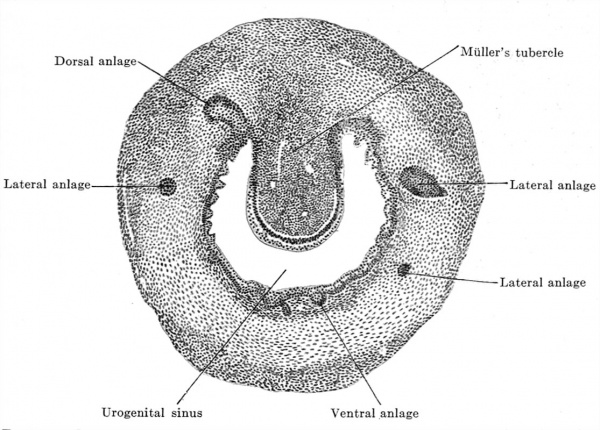
Fig. 655. Transverse section of the urogenital sinus of a male embryo of 70 mm. head-foot length. (Embryo R. Meyer 267, from the collection of Professor R. Meyer, Berlin; slide 60, row 2, section 3.) X47. The section shows Mailer's tubercle projecting into the lumen of the sinus and around the sinus the anlagen of the prostatic glands.
In its development the prostate belongs to the urethra as well as to the sinus urogenitalis, since the glands develop both above and below the opening of the primary excretory duct.
I have found the first anlage of the prostatic glands in a male embryo of 55 mm., and in a female embryo of 50 mm. It consists of solid epithelial buds that grow out into the surrounding thickened mesenchyme from the epithelium of the urogenital sinus (Figs. 628 and 655). These epithelial buds may arise around the whole periphery of the sinus, but they are most numerous on the dorsal surface, less so on the lateral surfaces and rare ventrally. Simultaneously with their appearance those of the dorsal and lateral surfaces branch, while those of the ventral surface remain unbranched and for the most part degenerate. The maximal number of glands that appear is twenty-six; a bilateral symmetry is unmistakable in the anlage. The direction of growth of all the glands is cranial, only the lowermost anlagen of the lateral surfaces grow caudally ; at feast two-thirds of the glands arise caudal to the openings of the primary excretory ducts.
The first muscle cells make their appearance in male embryos of 60 mm. head-foot length ; in the upper part of the prostate they are principally developed on the ventral surface, at the middle they form a closed ring and towards the lower border they are limited to the dorsal surface. Since the prostatic glands grow into this muscle mass, they force the muscle fibres apart and so produce the characteristic trabecular arrangement of the musculature. All the prostate glands are not united in the prostate, accessory glands occur in the interval between the prostate and Cowper's glands.
An embryo of 55 mm. head-foot length had four anlagen in the right side and none in the left; of those on the right 1 was above the primary excretoryduet and 3 below it. An embryo of 60 mm. head-foot length had 7 anlagen on the right side, 4 of which were dorsal and 3 lateral ; on the left there were 7 anlagen, of which 4 were dorsal, 2 lateral and 1 ventral. Of these anlagen 6 were above and 18 below the opening of the primary excretory duct. Another embryo of 60 mm. head-foot length had 11 anlagen on the right side, 7 of which were dorsal, 3 lateral and 1 ventral; on the left side there were 13 anlagen, 9 of which were dorsal, 3 lateral and 1 ventral. An embryo of 70 mm. head-foot length had 13 anlagen on the right side, 5 of which were dorsal, 5 lateral and 3 ventral; on the left there were 13 anlagen, 6 of which were dorsal, 4 lateral and 3 ventral ; 8 were above and 18 below the primary excretory duets.
In the female embryo few glands are formed, I have found as a maximal number only three ; they may undergo development and are then known as the paraurethral or Skene's ducts.
Development of the Bulbourethral (Cowper's) and the Vestibular (Bartholin's) Glands
Cowper's glands are formed from the pars pelvina of the urogenital sinus and are therefore of endodermal origin. Their first appearance has not been established. Lichtenberg (1906) found them already advanced in development in embryos of 48 mm. They arise as paired solid epithelial buds from the pars pelvina of the urogenital sinus. Frequently there are two on each side, and frequently one on one side and two on the other. The solid epithelial buds grow upwards almost parallel to the urogenital sinus, and lie from the beginning (Fig. 643) in the compact mesenchyme which forms the corpus cavernosum urethral The glands grow through this mantle, and only when they have reached the looser mesenchyme between the rectum and the sinus are they able to enlarge. Each gland will therefore possess an intrabulbar portion with few, short and slightly developed lateral branches, and an extrabulbar portion with numerous longer and richly branched lateral branches. The lateral branches appear immediately after the anlagen, the lumina develop in the main stem and in the primary branches in embryos of 65 mm., in the smaller later branches in those of 105 mm. (Lichtenberg 1906). In embryos of 120 mm. the transformation of the ordinary epithelium into a glandular epithelium takes place. The epithelium of the main stem also shows the character of glandular epithelium, and Lichtenberg (1906) is therefore right when he speaks, not of an efferent duct, but of a glandular stem tubule.
Bartholin's glands are the exact homologues of Cowper's glands, and, like them, arise as solid buds from the dorsal wall of the pars pelvina of the urogenital sinus. I first found them in an embryo of 36 mm. greatest length, their first division in one of 80 mm. head-foot length. The first appearance of a lumen in the efferent duct was observed by Spuler (1910) in an embryo of 82 mm. vertex-breech length, the first terminal vesicles in one of 120 mm., and the first formation of a secretion in embryos of from 150 to 160 mm. vertex-breech length. Up to and after birth there is a slow increase in the number of the terminal vesicles (Spuler 1910), and after puberty there is another more rapid growth (Huguier 1849). At the climacterium these glands undergo degeneration and may be entirely wanting in old age (Tiedemann 1840).
Development of the Small Sinus Glands
These glands also are of endodermal origin; only in the case of those situated in the glans penis is it impossible to exclude a. participation of the ectoderm. The glands appear in succession around the entire periphery of the sinus, first on the oral surface in embryos of 60 mm., on the right and left in embryos of 70 mm. and, finally, on the anal surface in embryos of 120 mm. (Lichtenberg 1906). These glands possess the same character as Cowper's glands, except that they do not attain to the same extensive development.
Descensus Testiculorum
It has been accepted by all authors as an established fact that the mesonephros, ovary and testis undergo an active displacement in the abdominal cavity. All three organs are supposed to gradually glide downwards from their original positions into the true pelvis, or, in the case of the testes, as far as the true pelvis. After what I have said as to the development of the mesonephros (p. 815 et seq.) and of the indifferent reproductive glands (p. 885 et seq.), it is clear that I cannot agree with this view. The descent of the mesonephros or the ovary and the internal descent of the testis to the inguinal canal does not take place, but on the other hand the testis does wander from the abdominal cavity into the scrotal sack. The anlagen of the mesonephros and of the reproductive glands extend from the diaphragm to or into the true pelvis. When the growth in length of the anlagen is completed their caudal ends become stationary, those of the mesonephros and testes at the boundary between the abdominal cavity and the primitive true pelvis, those of the ovaries in the true pelvis at the posterior surface of the genital cord. What seems to be a descent of the organs is caused, on the one hand, by their degeneration in their cranial portion, which produces a shortening of the entire organ and a downward displacement of its cranial end, and, in the second place, by the fact that the degeneration of the formed parts is proceeding while the anlagen themselves are still progressing caudally. We have to consider, then, only the external descent of the testis.
Between the testis, lying in the abdominal cavity, and the floor of the scrotal sack there is formed a band, the chorda gubernaculi.
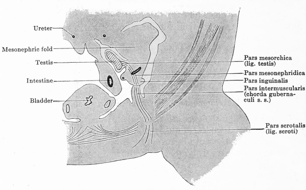
Fig. 656. The various parts of the*chorda gubernaculi are shown diagrammatically in a transverse section of the urogenital fold and body wall of a male embryo of 26 mm. greatest length. The chorda gubernaculi in the broader sense consists of 1, the lig. testis (pars mesorchica), 2, a mesenchymatous cord in the mesonephric fold and its plica inguinalis (pars mesonephridica), 3, a mesenchymatous cord in the crista inguinalis and between the abdominal muscles (pars intermuseularis), and 4, the lig. scroti (pars scrotalis).
whose development has already been discussed at two different places (p. 943), and may therefore be recapitulated here. Between the mesonephric fold and the lateral or anterior abdominal wall a connection is effected; the mesonephric fold sends out from its lateral surface a secondary projection, the inguinal fold; covered by a thickened epithelium and filled with a somewhat more compact mesenchyme; the lateral or anterior abdominal wall, on its part, forms an almost sagittal, ridge-like fold, the inguinal crest. The upper third of the crest lies opposite the fold, and in this upper third there develops in the crest a thickening of the mesenchyme, and spindle-shaped cells appear and form bundles that run in a horizontal direction from the crest towards the integument (Fig. 634). The fold and the upper third of the crest fuse together (Fig. 635), and thereby the connection between the mesonephric fold and the anterior abdominal wall is accomplished. Since the testis is united to the mesonephric fold by its mesorchium and by the epigonal portion of the genital fold, it too, along with the mesonephric fold, is attached to the anterior abdominal wall. The epigonal portion of the genital fold lies on the medial or dorsal side of the mesonephric fold, just where the plica inguinalis is given off from the ventral or anterior surface of the mesonephric fold (Fig. 635). We may, accordingly, recapitulate thus: The testis is connected with the anterior abdominal wall (1) by its mesorchium or the epigonal portion of the genital fold; (2) by the plica mesonephridica; (3) by the plica inguinalis, and, finally, (4) by the crista inguinalis (Fig. 656). Within the fold so constituted there is formed, at first in separate parts, a cord of closely compact, spindle-shaped cells, the chorda gubernaculi. In Fig. 656 the chorda has been inserted and its various parts are indicated by interruptions of the lines representing it; in the mesorchium and the epigonal portion of the genital fold the pars mesorchica develops, in the mesonephric and inguinal folds the pars mesonephridica, in the inguinal crest and the abdominal wall, so far as this is formed of the abdominal muscles, the pars intermuscularis, and, finally, in the integument, the pars scrotalis. This last streams out into the subcutaneous connective tissue of the genital swellings at- first, and into that of the unpaired scrotal area, when this is developed. In this way the caudal pole of the testis is connected with the testis sack and the chorda marks the path which the testis must traverse in its descensus.
The Formation of the Saccus Vaginalis
The saccus vaginalis is a part of the general body cavity that becomes completely cut off from the rest of the cavity by the formation of the anterior abdominal wall.
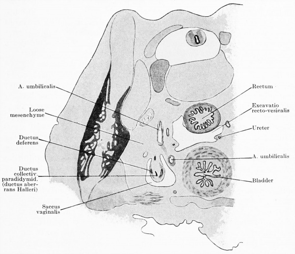
We may start the account of its development from the conditions seen in the body cavity of an embryo of 19.4 mm. greatest length (Fig. 631). The cavity may be divided into an upper broad portion and a lower narrow one, the primitive true pelvis. In the latter lie the rectum, projecting from the posterior wall, the urogenital fold projecting from the right and left walls, and, finally, the vesico-urethral anlage with the two art. umbilicales projecting from the anterior wall. At the boundary of the true pelvis and still lying in the primitive false pelvis is the union between the urogenital fold and the anterior abdominal wall (Fig. 631). Two processes bring about a change in these simple relations: (1) The bringing into the upright position of the anterior abdominal wall which in Fig. 631 is still horizontal, and (2) the taking into the body cavity of the loops of intestine that lie in the exoccelom. We may consider first the effect of this latter process. Space must be made in the body cavity for the loops of the intestines, which consist of almost the entire small intestine and a part of the large. This is accomplished by the enlargement of the sagittal diameter. The anterior abdominal wall thus becomes separated from the posterior one; but the mesonephric fold is attached to the anterior abdominal wall by the gubernaculum and the testis is attached to the mesonephric fold by the mesorchium and the epigonal portion of the genital fold. Both the mesonephric fold and the testis must therefore follow the anterior abdominal wall and become separated from the posterior one ; the caudal pole of the testis therebybecomes dircted ventrally and the long axrs changes from a vertical to a more horizontal position. The mesonephric fold always lies in front of the pole of the testis ; since it is attached not only to the posterior abdominal wall by its mesenterial portion, but also by its transition into the genital cord to the posterior wall of the vesicourethral anlage, it forms a loop around the testis, and the gubernaculum is attached at the summit of the loop. The formation of the loop naturally results in a stretching of the fold and its contents, and it is the medial limb of the fold that is most stretched, the lateral one, which forms the attachment to the posterior abdominal wall, being hardly stretched at all. The posterior abdominal wall follows the testis, an extensive layer of loose mesenchyme tissue becoming interposed between the ccelomic epithelium and the actual wall (Fig. 657). Thus the lower portion of the abdominal cavity, which in position corresponds to the fossa iliaca, is narrowed in its sagittal diameter to a third of its original size (Fig. 657), and appears to be a sack-like evagination of the undiminished portion of the abdominal cavity ; this sack-like portion is the anlage of the saccus vaginalis. What affects the mesonephric fold affects also its contents, but the primary excretory duct alone concerns us here. It also is thrown into a loop, whose summit corresponds to the first bend of the mesonephric fold, where the upper sagittal portion (Fig. 631) passes into the horizontal portion, and at this summit the tubuli collectivi of the paradidymis and, later, the ductus aberrans Halleri open (Fig. 657). It thus becomes certain that the entire ductus deferens is formed only from the medial limb of the loop of the excretory duct.
The bending of the anterior abdominal wall into the upright position will, of course, affect all the organs attached to its posterior surface; these are the bladder and the umbilical arteries. The bladder is simply brought from its bent position into an upright one, but it is different with the arteries. Since they arise from the aorta they are attached to both the anterior and posterior abdominal walls. From the aorta they run at first almost horizontally along the boundary between the false and true pelves (Fig. 631) to the anterior abdominal wall, and produce in this situation low peritoneal folds. Then they bend, and ascend towards the umbilicus along the bladder. When the arteries are drawn upwards by the pull exerted by the anterior abdominal wall, the horizontal portions are of course also raised, and by this elevation the peritoneal folds already present are materially increased in size.
Fig. 657. Part of a transverse section through a human embryo of 60 mm. head-foot length. (Embryo R. Meyer 264, from the collection of Professor R. Meyer, Berlin; slide 44, row 2, section 2.) By the union of the mesonephric fold and testis to the anterior abdominal wall, formed by the chorda gubernaculi, both have been carried quite away from the posterior abdominal wall. Between the urogenital fold and the posterior wall a layer of loose mesenchyme has formed. Thereby the portion of the abdominal cavity which contains the urogenital fold is separated off from the rest of the cavity as the saccus vaginalis. The separation between the two is completed by a peritoneal fold formed by the a. umbilicalis.
Rising from below like a septum, they form a partition between the primitive true pelvis and the anlage of the saccus vaginalis (Fig. 657), and the latter becomes marked off from the true pelvis and now appears to be a sack-like evagination of the body cavity, well defined on all sides, although actually, as we have seen, it is not an evagination, but a portion cut off from the general body cavity by the encroachment of the neighboring parts. In the region of the saccus vaginalis lie the caudal pole of the testis, the loop of the mesonephric fold and the gubernaculum. All three structures are at first so placed that they run through the centre of the saccus vaginalis, but later the lateral surfaces of the urogenital fold and gubernaculum fuse with the lateral wall of the. saccus, and the lumen of the saccus thus disappears on the lateral side and persists only on the medial one. The saccus vaginalis is so large that it extends both over and under the point of insertion of the gubernaculum into the anterior abdominal wall, that is to say, above and below the opening of the inguinal canal.
The so-called invagination of the saccus vaginalis into the abdominal wall is certainly at first not an invagination. The anterior abdominal wall thickens greatly, but the thickening does not affect the point at which the gubernaculum passes through the wall. Consequently the abdominal wall grows around the saccus and a groove appears in the wall for its reception (Fig. 635).
After this point there is a considerable hiatus in the history of the descent. We know that the saccus vaginalis eventually penetrates through the musculature, and, passing on below the former genital swelling, enters the scrotal sack. How this outgrowth takes place has not yet been determined. In the seventh month the testis wanders down through the inguinal canal and retains its original relation to the saccus, i.e., it is, as before, invaginated into it. The definitive position of the testis in the testis sack is acquired in the eighth month or at the latest before birth. The retention of a testis in the abdominal cavity or the inguinal canal is due to an inhibition of development, and constitutes what is known as cryptorchism.
After the completion of the descensus the saccus vaginalis remains connected with the abdominal cavity by a narrow canal. A short time after birth this becomes at first solid and is then resorbed. The saccus vaginalis, now completely cut off from the abdominal cavity, has become the tunica vaginalis propria testis. All possible variations in the extent of the degeneration of the narrow canal between the abdominal cavity and the tunica vaginalis propria may occur; parts of it may remain unclosed. If the opening of the canal into the abdominal cavity remains unclosed its persistence may predispose to an oblique inguinal hernia; a persistence of the entire vaginal canal may lead to the occurrence of a congenital inguinal hernia.
The tunica vaginalis communis develops from the fascia transversa. The spermatic fascia, as was pointed out above, is not a fascia but is formed from the hernia-like protrusion of the aponeurosis of the muse, obliquus abdominis externus.
In the female embryo there is an incomplete formation of a saccus vaginalis, which is frequently persistent as the diverticulum of Nuck, but otherwise it vanishes completely. The ovary cannot undergo a descensus similar to that of the testis because: (1) it descends lower than the testis; (2) it comes to lie in a different portion of the body cavity (the testis lies in the saccus vaginalis, the ovary in the recto-uterine excavation) ; (3) the lig. ovarii pro.prium is attached to the wall of the uterus, i.e., at the middle line.
Diagrammatic Representation of the Fate of the Mesonephros, Primary Excretory Duct, and Mullerian Duct in Both Sexes
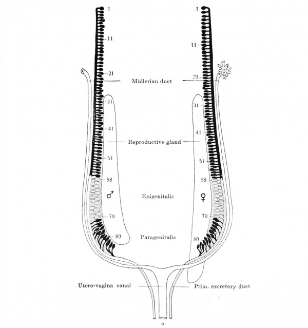
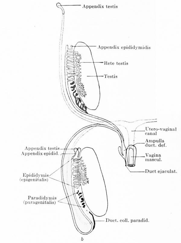
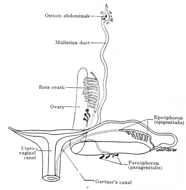
In conclusion I present in three diagrammatic figures (Fig. 658 a, b and c) a resume of the changes in position undergone by the organs of the urogenital fold in the two sexes. Those structures that are retained are represented by solid lines, and those that degenerate are outlined by broken lines ; the structures that change their position are shown in grey, and those that have attained their final position are in black. The diagrams show that no internal descent of the testis occurs, nor is there any downward migration of the ovary or mesonephros. The mesonephros and the reproductive gland are laid down throughout the entire length of the abdominal cavity, and their lower poles lie from the beginning at or in the true pelvis ; on the other hand, in both organs a cranial portion, which may include more than half of the entire organ, degenerates, and this degeneration may simulate a descent of the organs.
Fig. 658 a, b, and c. Diagrammatic representation of the entire renal organ, reproductive gland, and the efferent ducts. What persists is represented by solid, and what degenerates by dotted lines; whatever changes its position is shown in grey, whatever has reached its final position is in black, a. On the left is the male and on the right the female reproductive gland. Before the migration. The numbers are the serial numbers of the tubules.
Fig. 658 b. After the migration in the male embryo. The testis wanders out of the body cavity into the scrotal sack (descensus). The Mullerian duct degenerates throughout the greater part of its extent, the closed ostium abdominale persisting as the appendix testis (hydatid of the testis) , and the lowest portion of the utero-vaginal canal as the vagina masculina. All the tubules of the epididymis are not employed in the urogenital union: the first tubule remains free and becomes the appendix epididymis (hydatid of the epididymis) and the other unemployed tubules persist as appendices retis. The paradidymis (paragenitalis) is separated into its individual parts, some of the ductuli collectivi persist as the organ of Giraldes and the terminal portions of the ductuli, united to form a canal, persist as the ductus collectivus paradidymidis (ductus aberrans Halleri).
Fig. 658 c. After the migration in the female. The ovary remains in the body cavity, but rotates through 90° in such a way that its cranial pole comes to lie laterally, while the caudal pole, through which the axis of rotation passes, remains where it was and becomes the medial pole. The urogenital fold is rotated at the same time around the Mullerian duct, and as a result the tube comes to lie cranial to the ovary. The mesonephros and primary excretory duct degenerate ; of the mesonephros the epigenitalis persists as the epoophoron and parts of the paragenitalis as the paroophoron. Of the primary excretory duct only the portion that receives the tubules of the epoophoron is retained, though portions of the rest may persist as Gartner's canal.
The diagrams show furthermore that the tubules of the epigenitalis and paragenitalis — the persisting tubules of the mesonephros — are the most caudal tubules.
The testis undergoes an external descensus, during which no alteration of the relative position of the parts occurs. The ovary rotates through 90° and comes to lie with its long axis horizontal. Simultaneously with this rotation the genital fold rotates around its longitudinal axis, the Mullerian duct forming the axis. In consequence of this rotation, which is through almost 180°, the Mullerian duct comes to lie more cranially and the ovary more caudally.
The ostium abdominale of the Mullerian duct always lies in the neighborhood of the cranial pole of the ovary, notwithstanding that this wanders downwards as a result of the degeneration of the cranial portion of the ovary; this is due to the fact that its growth ceases for a considerable time, during which all the other organs increase materially in dimension.
Literature
In this list only the works cited in the text are included. The reader will find the remaining literature in the bibliography appended to the chapter on the Urogenital System in Hertwig's Large Handbuch, Vol. 3, or in the Normentafel of Keibel and Elze.
1 Since my manuscript was sent to the publisher, more than a year has elapsed. The literature has been brought up to July, 1911. although, with a few exceptions, only that which had appeared up to the summer of 1910 could be used in the text.
Ahlfeld, F. : Die Missbildungen des Menschen, F. W. Grunow, Leipzig, 18S0.
Alglave, P. : Note sur la situation du rein chez le jeune enfant par rapport a la crete iliaque et reflexions sur l'ectopie renale, Bull, et mem. Soc. Anat., vol. 85, p. 595-599, with 2 text-figs. Paris, 1910.
Biex, G. : Ueber Furchenbildung an der Oberflache des menschlichen Ovarium, Alonatsschr. f. Geburtsh. u. Gynakol., vol. 32, p. 175-179, with 1 plate. 1910.
Born, G. : Entwicklung der Ableitungswege des Urogenitalapparates und des Dammes bei Saugetieren, Anat. Hefte. Ergebnisse, vol. 3, 1894.
Boveri. T. : Die Niei'enkanalehen des Amphioxus. ein Beitrag zur Phylogenie des Urogenitalsystems der Wirbeltiere, Zool. Jahrb.. Abt. f. Ontogenie, vol. 5, p. 429-507, 1802.
Branca, Q. and Basseta, A. : Sur le developpement du testicule hmnaine, Arch.general de Chirurgie, vol. 1, p. 116-124, with 1 text-fig., 1907.
Braun : Ueber Abschniirung der Ovarien, Dissert, ined. Giessen, 1896.
Broek, van der, A. J. P. : Ueber den Schliessungsvorgang und den Bau des Urogenitalkanales (Urethra) beim menschlichen Embryo, Anat. Anz., vol. 37, p. 106-120, with 2 plates and 4 text-figs., 1910.
Buehler, A. : Beitrage zur Kenntnis der Eibildung beim Kaninehen und der Markstrange beim Fuchs und Menschen, Zeit. f. Wiss. ZooL, vol. 58, 1894.
Dandy WE. A human embryo with seven pairs of somites measuring about 2 mm in length. (1910) Amer. J Anat. 10: 85-109.
Delmas, J. and P. : Sur les anomalies ureterales, Ann. des malad. des Organes genitourin., vol. 28, 1, p. 865-896 and 984-1009.
Descomps: Anomalie des organes genitaux de la fern me. Arret de developpement. Uterus bicornis, ectopie de la trompe et de Fovaire, Bull, et Mem. Soc. Anat., Paris, vol. 84, p. 460-^61, with 1 text-fig., 1909.
DlSSE, J. : Untersuchungen fiber die Lage der menschlichen Harnblase und ibre Veranderungen im Laufe des Wachstums, Anat. Hefte, vol. 1, with 8 plates. 1891.
Elze, C. : Beschreibung eines menschlichen Embryo von ca. 7 mm. gr. L., Anat. Hefte, No. 106, with 7 plates and 32 text-figs., 1907.
Evatt EJ. A contribution to the development of the prostate in man. (1909) Jour. of Anat. and Phys. 43: 314-321.
Felix, W. : Die Entwicklung der Harnorgane, Hertwig's Handbuch der vergleich. u. experiment. Entwicklungsgesch., vol. 3, 1905.
Fleischmann, A. : Die Stilcharaktere am Urodaeum und Phallus, Morph. Jahrb., vol. 36, 1907.
Frankl, O.: Ueber Missbildungen der Gebarniutter und Tumoren der uterusligamente im Lichte embryologischer Erkenntnisse, Volkmann's Sammlung klinischer Vortrage, No. 363. 1902.
von Franque, O. : Ueber Umierenreste im Ovarium, zugleich ein Beitrag zur Genese der cystoiden Gebilde in der Umgebung der Tube, Zeit. f. Geburtsh. u. Gynak., vol. 49, p. 499-524, 1896.
Gebhard: Pathologisehe Anatomie der weiblicben Sexualorgane, Leipzig, 1899.
Gerard, L. : La forme de Furetere chez le fetus et le nouveau-ne, Medical Thesis, Paris, 1908.
Grubenmann, I. : Eine sagittale Verdoppelung der weiblicben Urethra, Inaug. Dissert. Zurich, with 2 plates and 5 text-figs., 1911.
Halban, J. : Die Grossenzunahme der Eier und Neugeborenen mit dem forsch reitenden Alter der Mutter. Arch. f. Entwieklungsmech., vol. 29, p. 439 455, 1910.
Hamburger, O.: Ueber die Entwicklung der Saugetierniere, Arch. f. Anat. u. Phys., Supplement, 1890.
Hart DB. The nature and cause of the physiological descent of the testes. (1909) J Anat Physiol. 43(3): 244-65. PMID 17232805
Hart DB. The nature and cause of the physiological descent of the testes. (1909) J Anat Physiol. 44(1): 4-26. PMID 17232824
Hart DB. The nature and cause of the physiological descent of the testes. (1909) Trans Edinb Obstet Soc. 1909;34:101-151. PMID 29612220
Hart DB. The physiological descent of the ovaries in the human foetus. (1909) J Anat Physiol. 44(1): 27-34. PMID 17232822
Hauch, E.: Ueber die Anatomie und die Entwicklung der Nieren, Anat. Hefte. vol. 22, p. 153-248, 1903.
Hegar K. Anatomical investigations on the nullipara uterus with special consideration of isthmus (Anatomische Untersuchungen am nullipara Uterus mit besonderer Berücksichtigung der Isthmus). (1908) Beitraae zur Geburtsh. u. Gynak., vol. 13, 1908.
Herzog, F. : Beitrage zur Entwicklungsgeschichte und Histologie der miinnlichen Harnrohre, Arch. f. mikr. Anat. u. Entw., vol. 63, 1904.
Hochstetter, F. : Ueber die Arterien des Darmkanales der Saurier, Morph. Jahrb., vol. 26.
Holzbach, E. : Die Hernmungsbildungen der MUller'schen Gange im Lichte der vergleiehenden Anatomie und Entwicklungsgeschichte, Beitriige zur Geburtsh. u. Gynak., vol. 14, p. 3-57, with 6 text-figs., 1909.
Huguier : Memoire sur les appareils secreteurs des organes genitaux externes chez la femme et chez les aniruaux, Ann. des Sci. nat., Series 3, vol. 13, 1849.
Ingalls NW. Description of a human embryo of 4.9 mm (Beschreibung eines menschlichen Embryos von 4.9 mm). (1907) Arch. f. mik. Anat., 70: 506-576.
Ingalls, N. W. : Beschreibung eines rnensehlichen Embryo von 4.9 mm. L., Arch. f. mikr. Anat. u. Entw., vol. 70, p. 506-570, with 3 plates and 28 textfigs., 1907.
Jixsabato, L. : Sul connetivo nell' utero fetale con particolare riguardo alia sua istogenesi, Monit. Zool. Ital., vol. 19, p. 281-285, 1908.
Kehrer, E. : Das Nebenhorn des doppelten Uterus, Inaug. Dis., Heidelb., 1899.
Keibel, F. : Zur Entwicklungsgeschichte des mensehlichen Urogenitalapparates, Arch. f. Anat. u. Entw., 1896.
Kolliker, A. : Lehrbuch der Entwicklungsgeschichte, 2nd ed., Engelmann, Leipzig, 1879.
Kossmanst, R. : Missbildungen und Lageanomalien, in Martin, Krankheiten der Eileiter, Leipzig, 1895.
Kosshann, R. : Mangel, Unvollkommenheit, Ueberzahl, Verlagerung der Eierstocke, in Martin, Krankheiten der Eierstocke, Leipzig, 1899.
Krieger: Monatsschr. f. Geburtsk., vol. 12, p. 179, 1858 (cited by Kussmaui, 1859).
Kuelz, L. : Untersuchungen iiber das postfetale Wachstum der mensehlichen Niere. Inaug. Dissert. Kiel, 1899.
Kussmaul, A. : Von dem Mangel, der Verkümmerung und Verdopplung der Gebärmutter (Of the defect of atrophy and duplication of uterus), Wiirzburg, 1859.
Lams and Doorme : Nouvelles recherches sur la maturation et la f eeondation de l'oeuf des mammiferes, Arch, de Biologie, 1907.
Legueu, F. : L'anatomie chirurgicale du bassinet et l'exploration interne du rein, Ann. de Gynec, 1891.
Leydig, F. : Lehrbuch der Histologie des Menschen und der Tiere, Frankfurt a. M., 1857.
Lichtenberg, A. : Beit rage zur Histologie, mikroskopischen Anatomie und Entwicklungsgeschichte des Urogenitalkanales des Mannes und seiner Driisen, Anat. Hefte, vol. 31, 1906.
Menge, C. : Bildungsfehler der weiblichen Genitalien, in Veit's Handbuch der Gyniikologie, J. F. Bergmann, Wiesbaden, 1910.
Meves and Duesberg : Ueber Mitochondrien bzw. Chondrioconten in den Zellen junger Ernbryonen, Anat. Anz., vol. 31, 1907.
Meves and Duesberg : Die Chondriosomen als Trager erblicher Anlagen, Arch. f. mikr. Anat. u. Entw., 1908.
Meyer, R. : Ueber die fetale Uterusschleimhaut, Zeit. f . Geburtsh. u. Gynak., vol. 38, 1898. Meyer, R. : Zur Entstehung des doppelten Uterus, Zeit. f . Geburtsh. u. Gynak., vol. 38, 18986.
Meyer, R. : Ueber epitheliale Gebilde im Myometrium des fetalen und kindlichen Uterus einsehl. des Gartner'schen Ganges, Berlin, 1899.
Meyer, R. : Ueber sogenannte Vornierenreste und das nephrogene Zwischenblastem bei mensehlichen Embryonen und ihre eventuelle pathologische Persistenz, Charite-Annalen, vol. 33, p. 649-656, with 2 text-figs., 1909.
Meyer, R. : Zur Kenntnis des Gartner'schen Ganges besonders in der Vagina und dem Hymen des Menschen, Arch. f. mikr. Anat., vol. 73, 1909.
Meyer, R. : Zur Entwicklungsgeschichte und Anatomie des utrieulus prostaticus beim Menschen, Arch. f. mikr. Anat. u. Entw., vol. 74, 1909b.
Meyer, R. : Ueber erosio portionis uteri, Deut. patholog. Gesellsch. Erlangen, 1910.
Meyer, R.: Die Erosion und Pseudoerosion des Erwachsenen, Arch. f. Gynak. vol. 91, part. 3, p. 1-35 with 1 plate and 3 text-figs., 1910.
Meyer, R. : Die Epithelentwicklung der cervix und portio vaginalis und die pseudoerosio congenita (Kongenital. histol. Ectropium), Arch. f. Gynak., vol. 90, part 3, p. 1-20 with 1 plate, 1910.
Meyer, R. : Beitrag zur Frage nach der Genese der im Urogenitalgebiet vorkommenden Mischgeschwiilste und Teratome, Reprint from the Charite Annalen, vol. 34, p. 1-23 with 10 text-figs., 1910.
Nagel, W. : Ueber die Entwicklung des Uterus und der Vagina beim Menscken, Arch. f. mikr. Anat., vol. 36, 1891.
Nagel, W. : Entwicklung und Entwicklungsfehler der weiblichen Genitalien, in Veit's Handbuch der Gynak., J. F. Bergmann, Wiesbaden, 1897.
Ogushj, K. : Zur Frage des mensehlichen Eidotters, Anat. Anz., vol. 37, p. 83-86 with 1 fig.
Papin, E. : Contribution a, Petude des anomalies de Puretere, duplicite et bifidite des ureteres (1. mem.), Revue de Gynecol., vol. 15, p. 108-132 with 26 figs., 1909.
Perna, G. : Sullo sviluppo e sul significato delP uretra cavernosa delP uomo, Soc. med. chir. di Bologna Adunauze, 1909.
Perna, G. : Sullo sviluppo e sul significato delP uretra nelP uomo, Arch. Ital. Anat. e EmbrioL, vol. 8, p. 145-154, 1909.
Peter, K. : Untersuchungen fiber Bau und Entwicklung der Niere. I. Die Nieren kanalchen des Menschen und einiger Saugetiere, G. Fischer, Jena, 1909.
Pick, L. : Gebarmutterverdoppelung und Geschwiilstbildung unter Berucksichtigung ihres atiologischen Zusamrnenhanges, Arch. f. Gynak., vol. 52, 1896.
Pick, L. : Zur Anatomie und Genese der doppelten Gebarmutter, Arch, f : Gynak., vol. 57, 1898. Piquand, G. : Les uterus doubles. Anatomie et developpement, Revue de Gynecol., vol. xv, p. 401-466, 1910.
Popoff, N. : Zur Morphologie und Histologie der Tnben und des Parvoariums beim Menschen wahrend des intra- und extrauterinen Lebens bis zur Pubertat, Arch, f . Gynak., vol. 44, 1893.
Popoff, N. : L' ovule male et le tissu interstitiel du testicule chez les animaux et ehez Phomme, Arch, de Biologie, vol. 24, p. 433-500 with 3 plates, 1909.
von Recklinghausen: Die Adenomyome und Cystadenome des Uterus und der Tubenwandungen, ihre Abkunft von Resten des Wolffschen Korpers, Berlin, 1896.
Reichel, P. : Die Entwicklung der Harnblase und der Harnrohre, Verb. phys. med. Gesellsch. Wiirzburg, 1893.
Riedel: Entwicklung der Saugetierniere, Unters. anat. Inst. Rostock, 1874. Rielander, A.: Das Paroophoron (vergl. anat. u. path. -anat. Studie), Marburg, 1904.
Roesger., P. : Zur fetalen Entwicklung des mensehlichen Uterus, insbesondere seiner Muskulatur, Festschr. z. 50 jahr. Jubil. d. Gesellisch. Geburtsh. u. Gynak., Berlin, 1894.
Romanowsky, R. and von Winiwarter, J. : Dystopia testis transversa, Anat. Anz., vol. 26, 1905.
Rubaschkin, W. : Ueber die Urgeschlechtszellen bei Saugetieren, Anat. Hefte, No. 119 (vol. 39), p. 603-652 with 4 plates and 6 text-figs., 1909.
Rubaschkin, W. : Chondriosomen und Differenzierangs-prozesse bei Saugetier embryonen, Anat. Hefte, No. 125 (vol. 41), with 4 plates and 15 textfigs., 1910.
Rudolph : Eine Hemmungsbildung weiblicher Geschlechtsorgane, Med. Dissert., Bonn, 1909.
Schoener, O. : Bestimmung des Geschlechtes am mensehlichen Ei vor der Befruchtung und wahrend der Schwangerschaft, Beitrag z. Geburtsh. u. Gynak., vol. 14, p. 454-475, 1909.
Schreiner, K. E.: Ueber die Entwieklung der Amniotenniere, Zeit. f. wiss. Zool., vol. 71, p. 1-188, 1902.
Schweigger-Seidel : Die Nieren des Menschen und der Siiugetiere in ihrem feineren Ban, Halle, 1865.
Seitz, L. : Ueber die Form der Ureteren, speziell bei Feten nnd Nengeborenen, Beitr. z. Geburtsbilfe u. Gyniik., vol. 13, with 5 figs., 190S.
Spuler, A. : Ueber die normale Entwieklnng des weiblichen Genitalapparates, Veit's Handbuch der Gyniik., vol. 5, J. F. Bergmann, 1910.
Stoerk 0. Contribution to the knowledge of the structure of the human kidney (Beitrag zur Kenntnis des Aufbaus der menschlichen Niere). (1904) Anat. Hefte, No. 72.
Stricht, van der : La structure de l'oeuf des mammif eres. II Partie, Bull, de l'Acad. roy. de med. de Belgique, 1900.
Stricht, van der: La structure de l'oeuf des mammif eres. Ill Partie, Mem. publ. par la classe des Sciences de l'Acad. roy. Belgique, 1909.
Tandler, J. : Ueber Vornierenrudimente beim menscblicben Embryo, Anat. Hefte, 1905.
Tandler, J. : Ueber den Einfluss der innersekretoriscken Anteile der Geschlechtsdriisen auf die aussere Erscbeinung des Menschen, Klin. Wochenschr., vol. 23, No. 13, p. 1-24, Vienna, 1910.
Tausig, F. J. : Die Entwieklung des Hymen, Monatsscbr. f . Geburtsh. u. Gyniik., vol. 30, p. 696^705 with 13 text-figs., 1909.
Thiersch, C. : Bildungsfehler der Ham- und Geschlechtswerkzeuge des Mamies, Ulustr. Med. Zeitschr., vol. 2, 1S52.
Tiedehann, Fr. : Von den Duverney'schen, Bartholini'schen oder Cowper'schen Driisen des Weibes, Heidelberg and Leipzig, 1840.
Toldt, C. : Untersuchungen liber das Waehstum der Nieren des Menschen und der Siiugetiere, Sitzungsber. Akad. Wissensch. YVien, Abt. Ill, vol. 69, 1874.
Tourneux, J. : Sur les premiers developpements du tubercle genitale etc. chez le mouton, Comptes rendus Soc. biol., ser. 8, vol. 5, 1888.
Tourneux, J. and Legay : Developpement de l'uterus et du vagin depuis la fusion des conduits de Midler a la naissance, Comptes Rendus Congr. internat. sciences med., Copenhagen, 1887.
Tschaschin, S. : Ueber die Chondriosomen der Urgeschlecbtszellen bei Vogel embryonen, Anat. Anz., vol. 37, with 8 text-figs., 1910.
Veit, O. : Ueber das Vorkommen von Urnierenrudimente und ihre Beziehungen zur Urniere beim Menschen, Marburger Sitzungsber., 1909.
Versari, R.: La morfogenesi della guaina dell' uretere umano, Atti R. Acad. Sci. Med., 1909; Palermo, 1910.
Waldeyer, W. : Eierstock und Ei, Engelmann, Leipzig, 1870.
Weber. S. : Zur Entwicklungsgeschichte des uropoetischen Apparates bei Saugern, mit besondei-er Beriicksichtigung der Urniere zur Zeit des Auftretens der bleibenden Niere, Dissert, med., Freiburg i. Br.. 1892.
Werth and Grusdew : Ueber die Entwieklung der menschlichen Uterusmuskulatur. Arch. f. Gyniik., vol. 55, 1898.
Wimmer, H. : Doppelbildungen an den Nieren und ein Versuch ihrer entwicklungsgescliichtlichen Deutung, Virchow's Arch., vol. 200, p. 487-522 with 14 figs., also as Dissert, med. Munich. 1910.
von Winckel : Ueber die Einteihrag, Entstehung und Benennung der Bilden?s hemmungen der weiblichen Sexualorgane, Volkmann's- Sammlung klin. Vortrage, 251-252, Leipzig, 1899.
von Winiwarter, II.: Recherches sur l'ovogenese el l'organogenese de l'ovaire des mammif eres (Lapin et Homme), Arch, de Biol., vol. 18, p. 33-199 with 6 plates, 1900.
Wyder, Th. : Beitriige zur normalen und pathologischen Histologie der menschlichen Uterusschleimhaut, Arch. f. Gvniik.. vol. 13, 1S7S.
| Historic Disclaimer - information about historic embryology pages |
|---|
| Pages where the terms "Historic" (textbooks, papers, people, recommendations) appear on this site, and sections within pages where this disclaimer appears, indicate that the content and scientific understanding are specific to the time of publication. This means that while some scientific descriptions are still accurate, the terminology and interpretation of the developmental mechanisms reflect the understanding at the time of original publication and those of the preceding periods, these terms, interpretations and recommendations may not reflect our current scientific understanding. (More? Embryology History | Historic Embryology Papers) |
Cite this page: Hill, M.A. (2024, April 27) Embryology Book - Manual of Human Embryology 19-4. Retrieved from https://embryology.med.unsw.edu.au/embryology/index.php/Book_-_Manual_of_Human_Embryology_19-4
- © Dr Mark Hill 2024, UNSW Embryology ISBN: 978 0 7334 2609 4 - UNSW CRICOS Provider Code No. 00098G

