Book - Manual of Human Embryology 1
| Embryology - 27 Apr 2024 |
|---|
| Google Translate - select your language from the list shown below (this will open a new external page) |
|
العربية | català | 中文 | 中國傳統的 | français | Deutsche | עִברִית | हिंदी | bahasa Indonesia | italiano | 日本語 | 한국어 | မြန်မာ | Pilipino | Polskie | português | ਪੰਜਾਬੀ ਦੇ | Română | русский | Español | Swahili | Svensk | ไทย | Türkçe | اردو | ייִדיש | Tiếng Việt These external translations are automated and may not be accurate. (More? About Translations) |
Keibel F. and Mall FP. Manual of Human Embryology I. (1910) J. B. Lippincott Company, Philadelphia.
| Historic Disclaimer - information about historic embryology pages |
|---|
| Pages where the terms "Historic" (textbooks, papers, people, recommendations) appear on this site, and sections within pages where this disclaimer appears, indicate that the content and scientific understanding are specific to the time of publication. This means that while some scientific descriptions are still accurate, the terminology and interpretation of the developmental mechanisms reflect the understanding at the time of original publication and those of the preceding periods, these terms, interpretations and recommendations may not reflect our current scientific understanding. (More? Embryology History | Historic Embryology Papers) |
Keibel F. I. The Germ Cells in Keibel F. and Mall FP. Manual of Human Embryology I. (1910) J. B. Lippincott Company, Philadelphia.
I. The Germ-Cells
By Franz Keibel Freibueg i. Bb.
The germ-cells of man are the ovum (oide, avium, mature ovum), formed in the ovary, and the spermion, formed in the testes. The mature human ovum (the oide of Korschelt and Heider, the avium of Waldeyer) is not yet known, nor have the processes which bring about its maturation yet been certainly observed. Nagel[1](1888) in connection with his PI. XXI, Fig. 7, speaks of remains of polar bodies, but this interpretation, as Waldeyer (1902, in Hertwig's Handbuch, vol. i, part i, p. 333) has pointed out, cannot be accepted in view of the occurrence in the ovimi figured of a perfectly unaltered nucleus.
The human ovmn which has reached its full size in the ovary is a true cell, with cytoplasm, in the ovum termed ooplasm (yolk), a nucleus, frequently spoken of as the germinal vesicle, and a nucleolus, also termed the germinal spot. In the nucleus there is, in addition to the nucleolus, a fine, somewhat scanty nuclear reticulum containing chromatin. We must assume that processes of maturation occur in such an ovum, similar to those which occur in the ova of other animals. Briefly stated the maturation consists in that from the ovum, by two rapidly succeeding divisions, four cells, one large and three small, are formed. The large cell is the mature ovum, the oide or ovium, the three other cells are the polar bodies (polocytes), formerly known as the directive corpuscles. At the first division the first polar body is separated, at the second division the second one, the first one at the same time also undergoing division. The divisions take place by mitosis and the final products possess only half the number of chromosomes characteristic of the ordinary cell-divisions of the species. A reduction of the number of chromosomes by one-half is thus brought about, but how this reduction is accomplished and what may be its significance is still a matter of discussion; indeed, whether the reduction always actually takes place during the nuclear divisions which produce the polocytes — whether at the one or the other of these divisions there is really a reduction division — is yet imcertain. For further consideration of this point reference may be had to Korschelt and Heider,[2] Haecker,[3] Waldeyer,[4] and E. B. Wilson.[5] Individually many variations occur in the process; the division of the first polocyte may not take place, the formation of one of the polar bodies may be suppressed, and, what is noteworthy, modifications occur in individuals of one and the same species.
The centrosome of the egg-cell seems usually to degenerate during these processes. The time, with reference to the fertilizaticm, at which the formation of the polar bodies takes place, varies; the first one is often formed while the ovum is still in the ovary. In mammals these phenomena have been especially well studied in the mouse (Sobotta,[6] Leo Gerlach,[7] and others), and they have also been investigated in the guinea-pig (Rubaschkin). In man, as has been stated, nothing is known of these things. We may with perfect certainty assume that polocytes are formed and that there is a reduction of the chromosomes to one-half the typical number; but whether variations from the general plan occur, whether or not the formation of one of the polar bodies is suppressed, we do not know. Similarly we can say nothing as to the time at which the polocytes are formed. It may be that the first one is formed while the ovum is still in the ovary, and observations on this point are much to be desired.
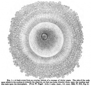
But even although, as is clear from what has just been said, the mature human ovum is unknown, nevertheless descriptions have frequently been given of a so-called mature human ovum, that is to say, of an ovum which was near maturity, and usually the figure and description given by Nagel has formed the basis of these accounts. 0. Hertwig follows Nagel's description and Waldeyer quotes it almost verbally ; it may be given here as well as the figure.
The human ovum retains its transparency in all stages of development, so that all its anatomical characteristics can be fully made out in the living object. The cell substance, which may be termed the ooplasma, and which is frequently spoken of as the yolk, is separated into two layers. In the inner (central) layer is found the greater part of the deutoplasm, that is to say, the inclusions of the ovum, which are usually regarded as nutritive or reserve substances; they produce merely a slight opacity in comparison with what is found in the ova of other mammals. The deutoplasm consists partly of feebly and partly of strongly refractive, finer and coarser granules; but a distinct delimitation of the deutoplasmic elements, such as one finds not only in the ova of the lower vertebrates but also in those of many mammals, cannot be made out. The outer layer, the marginal zone of the ooplasma, is much more finely granular and transparent. It contains the germinal vesicle, that is to say, the nuclens of the cell, in which is to be seen a large germinal spot or nacleolus. In ova examined while fresh in the liquor follieuli Nagel observed amoeboid movements in the nncleolus, and such movements have also been described as occurring in the nucleoli of other ova, although Flemming ("Zellsubstanz, Kern, und Zellteilung," Leipzig, 1882, p. 157), to whom Nagel (*'Die weibliche Geschlechtsorgane," in K. von Bardeleben's "Handbuch der Anatomie des Menschen," vol. vii, p. 59, 1896) refers in connection with the egg of Paludina, expressly states that in spite of many endeavors he had not succeeded in perceiving changes of form in living nucleoli. Further observations on this point are much needed. The nucleus appears to be homogeneous in the freshly stained ovum, but with proper staining a scanty chromatin network becomes visible. The membrane which encloses the ovum, the zona pellucida, is remarkably broad and is finely striated radially. It is separated from the ooplasma by a narrow perivitelline space and is surrounded by cells, derived from the cumulus ovigerus which surrounds the ovum in the ovarian follicle.
These cells form three or four layers, those of the layer nearest to the zona pellucida having a markedly radiating arrangement and forming in the fully formed ovum the corona radiata of Bischoff. This author regarded the corona as an indication of the maturity of the ovum, a view with which Waldeyer coincides to the extent that he regards a well-developed corona as a sign of approaching maturity, without, however, acknowledging it to be an indication of complete ripeness. In this he agrees with Van Beneden and Nagel.
To this description of the approximately ripe ovum it must be added that the existence of a perivitelline space is in dispute. Nagel believes that the space is of importance in that it permits a rotation of the ovum so that the nucleus usually lies upon its upper surface, for this was the position in which he found it in all fresh, approximately ripe ova. Ebner (Kolliker's "Handbuch der Gewebelehre," vol. iii, p. 517, 1902) disputes both the existence of a perivitelline space and the assumption that the ovum rotates witliin the zona pellucida, the nucleus thereby coming to lie upon the upper surface. He is of the opinion that the nucleus ascends through the almost fluid ooplasma on account of its lesser specific gravity, and that the ovum as a whole does not rotate; he bases his opinion on the fact that when a fresh ovum is burst the greater part of the ooplasma together with the nucleus is expelled, but the apparently denser marginal zone of the ooplasma always remains adherent to the zona pellucida. This could not occur if an interval jfiUed with fluid was interposed between the marginal ooplasma and the zona. Furthermore it could be observed, by carefully focussing an equatorial optical section of an uninjured fresh ovum, that the marginal ooplasma was in intimate contact with the inner surface of the zona pellucida. **What Nagel figures as a cleft is a line of refraction, which, with deep focussing, appears to be in the deeper layer of the yolk (ooplasma), and apparently separates the yolk (ooplasma) and the zona, but is really a purely optical phenomenon which depends on the curvature of the zona and resembles the line of refraction which one may observe under similar circumstances in cartilage cavities."
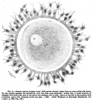
Waldeyer (l. c, p. 332), on the other hand, mantains the correctness of Nagel's observation, since he has been able to convince himself of it from the ovum figured by Nagel. He is not, however, convinced as to the rotation of the ovtmi within the zona, but agrees with Ebner that the fact observed by Nagel may be explained by the ascent of the lighter nucleus through the almost fluid ooplasm. He himself shows in his Fig. 134 (L c), which is reproduced here as Fig. 2, an almost ripe human ovum (a fully grown, oocyte), taken from an ovary while it was still warm, in which there was not the slightest indication of a perivitelline space. Waldeyer explains this discrepancy by assuming that the ovum described by Nagel was farther advanced towards maturity. The ooplasm is also ai-ranged somewhat differently in this ovum from what it is in that described by Nagel. Close under the zona there is a very narrow zone of a finely granular material, the ooplasm cortex, which was clearly seen by Waldeyer as well as by Ebner; beneath this is a broader, clearer zone of ooplasm in which the nucleus lies in almost ripe ova, and then comes the central darker mass of the ooplasm.
It will be seen that in this connection important questions still await decision, and if gynaecologists and embryologists work with a common purpose a solution of them may be expected. Another question I regard as already settled. The human ovum has no micropyle, that is to say, no preformed opening in the zona pellucida for the entrance of the spermium. The question as to the existence of a micropyle has been recently revived by Holl {Anat Am., 1891, p. 554; and Sb. K. Acad. Wien., vol. cii, 1893), after it had long been regarded as settled. It seems to me to be certain that Holl has been the victim of a mistake. Ebner (Kolliker's * * Gewebelehre, " vol. iii, p. 518) says **Holl's figure of a section of an apparently degenerated human ovum shows, traversing the greatly shrunken zona, a small oblique cleft, probably formed by accident, perhaps by a wandering cell. True micropyles, so far as known, traverse the zona radially, not obliquely."
The development of the ovum will not be discussed here, but in the chapter on the urogenital apparatus; yet, since it has to do with the question of the relation of the follicle cells to the ooplasm, it must be noted here that the mode of formation of the zona pellu cida is not yet certain. Waldeyer inclines to the opinion that it is a product of the ooplasm and is therefore a primary egg-m.embrane. E. Hertwig (0. Hertwig's *'Handbuch") and others regard it as a chorion, that is, as a secondary egg-membrane, a product of the follicle cells; while for others it is a double product of the follicle cells and the ooplasm, at least so Waldeyer interprets the observations of Retzius,[8] Flemming,[9]and Ebner.[10] According to these authors there is first formed, from processes extending from the follicle cells to the ooplasm, a delicate network, which rests closely upon the surface of the ovum. The network is the first indication of the zona, and, later, a homogeneous substance appears between its fibres and forms with them the zona. Whence this homogeneous substance arises is uncertain; according to Waldeyer it may come from the ooplasm. A portion of the cell bridges tliat originally extend between the follicle cells and the ooplasm are retained in the forming zona substance in a protoplasmic condition, but whether such unions persist in the approximately ripe ovum is uncertain ; certainly their existence can hardly be reconciled with the presence of a perivitelline space. Kolliker (according to Von Ebner in Kolliker's Handbuch der Gewebelehre," vol. iii, p. 511) gives the diameter of the approximately ripe ovum as 0.22-0.32 mm., but Waldeyer remarks {I. c, p. 352, note) that he has never seen a human ovum of over 0.25 mm. As regards further measurements Ebner gives the diameter of the nucleus (germinal vesicle) as 30-45 fi, that of the nucleolus (germinal spot) as 7-10 /A, that of the zona pellucida as 10-11 /a, and that of the deutoplasm granules (yolk granules. Von Ebner) as 2-3 II.
The production of ova begins at a very early period of life in the human species. According to Waldeyer (l. c, p. 373; and "Eierstock und Ei," 1870) at birth or shortly thereafter all the oogonia have become oocytes of the first order, and so have before them only further growth and maturation. (The contrary opinion of Paladino[11] I do not consider well founded.) Already in the ovary of the child the ova may approach ripeness (see C. Hennig; Ueber fruhreife Eibildung, Sb, d. Leipzig, Naturf. Ges., p. 5, 1878). Also Waldeyer says, One finds in the ovaries of newly-born and young children follicles the size of a pea with normally developed ova." On the other hand, those ova which ripen only after the cessation of ovulation require for their development about fifty years.
Into the broad field of the pathology of the ovum I cannot enter here; nevertheless, follicles with several ova, multinucleated ova, and the fragmentation of the ovum may be briefly mentioned. Follicles with several ova may be explained (Schottlander: Arch, f, mikr. Anat,, vol. xli, 1893; M. and P. Bouin; C, R, Soc. BioLy vol. lii, p. 17 and 18, Paris, 1900 [dog] ; Ch. Honore: Arch, de Bid., vol. xvii, p. 489-497, 1900 [rabbit] ) by supposing that the different ova of an egg mass in the embryo and child have not completely separated, so that several ova have become enclosed within a common follicle wall. They may very naturally tend to the production of twin pregnancies. They may also be supposed to have arisen by the fusion of originally distinct follicles. Multinucleated ova have been accounted for in various ways and perhaps have various methods of origin. They may be formed by direct nuclear division (Stockel: Arch. f. mikr. Anat., vol. liii, 1899; Falcone: Monitors zool. ital., Suppl., 1899) or one may suppose that originally distinct ova have subsequently fused (H. Rabl: Mehrkemige Eizellen und mehreiige Follikel, Arch, f, mikr. Anat., vol. liv, 1899; S. von Schumacher und C. Schwarz: Anat. Anz.y vol. xviii, 1900). Finally, they may be produced by the division of the nucleus of an oogonium, without the corresponding division of the cytoplasm taking place, as sometimes occurs in spermatogonia. Cases in which several nuclei occur in an ovum as the result of an imimigration of leucocytes need not be considered here.
In mammals a division or fragmentation of ripe ova after the expulsion of polar bodies has been observed (Henneguy, Janosik, H. Rabl, Gurwitsch, Van der Stricht), and I have also seen such a condition in human ova. The phenomenon is one leading to the degeneration of the ovum; some authors have compared it with segmentation and have seen in it a parthenogenetic process, but Bonnet[12] has disposed of such notions.
We may now turn to a consideration of the male germ-cell, the spermium, which is formed in the testis. Tt is well known that for a considerable time it was uncertain whether the male cells were not parasites in the seminal fluid — the name spermatozoa is a reminder of this idea. The development of the spermium first -. I clearly showed that this structure is nothing else
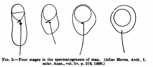
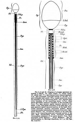
Spermatogenesis has also been studied in man and some figures from Meves[13] {Arch. f. mikr. Anat., vol. liv, p. 378) may I be reproduced here. Other figures have been I given by Ebner (Kolliker's Handbuch," vol. iii, p. 454). In the study of human spermatogenesis attempts have been frequently made to determine the number of the chromosomes. Duesberg,[14] who also cites the literature bearing on the question, finds that in the spermatocytes there are in all probability twelve. If this be correct in the spermatogonia and the soma cells there would be twenty-four, as Flemming had already (1898) supposed. Good figures of human spermia have recently been given by Betzius {Biol. Unters., neue Folge x, 1902), and an excellent diagram by Meves is reproduced by Waldeyer in Hertwig's Handbuch," vol. i, p. 146. In the human spermium, which is essentially similar to that of other mammals, there may be recognized a head and a tail; a neck piece is not clearly distinguishable. In the tail, if the indistinct neck piece be disregarded, there are a connecting piece, a principal piece, and an end piece. Seen from the surface the head is oval, and in side view it is elongated pear-shaped, the tail being attached to the broader end ; upon each surface of the head there is a slight depression.
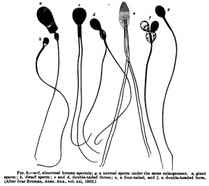
According to Waldeyer one sees with very strong magnification a constriction between the head and the connecting piece, and this is an indication of the neck; and in this situation Krause and Waldeyer describe a small depression in the head, which receives the neck together with the connecting piece. By staining the head cap can be brought into view, its posterior border marking off an anterior and a posterior portion of the head. The anterior sharp border of the cap represents the perforatorium; special perforatoria, such as Nelson[15] and Bardeleben[16] have described, may be produced by special conditions and perhaps have been confused with attached bacteria. The neck has the form of a disk, which is formed by the anterior centrosome bodies, the noduli anteriores (Fig. 5, A and B, Nd.a, dark), and a homogeneous intermediate substance, the massa intermedia {Ms.int., clear). The succeeding connecting piece (pars conjunctionis) begins with the noduli posteriores (the posterior centrosome bodies), represented in the diagram as a black stripe, and ends with the annulus ; it includes, therefore, the region of the posterior centrosome, which during spermatogenesis has divided into these two portions. The filum principale of the tail traverses the axis of the connecting piece, extending from the proximal portion of the posterior centrosome. In this region the filum principale has a delicate investment which probably passes over posteriorly into the thicker sheath of the tail and finally ends at the beginning of the filum terminale. Around this delicate sheath is the spiral sheath, and external to this the mitochondria sheath. The spiral sheath consists of a spiral filament, not recognizable in the mature spermium, and an intermediate substance, the substantia intermedia, represented as clear in the diagram. The mitochondria sheath is the matrix of the spiral filament and is characterized by the presence of mitochondria granules. At the beginning of the principal portion of the tail the spiral and mitochondria sheaths terminate, but the inner thin sheath is probably continued into the involucrum of the tail. A spiral membrane has been described for the human spermium by several authors, but does not really occur. The measurements of the human spermium are, according to W. Krause ("Handbuch der menschl. Anat.," vol. i, p. 559, 1876), as follows : Entire length 52-62 fi, of which the head measures 4.5 /*, the connecting piece 6 /a, and the tail 41-52 /a. The width of the head is 2-3 microns its thickness 1-2 microns. Giant spermatozoa also occur. Such a structure was figured by Widersperg[17] in 1885. Since then abnormal forms of human spermatozoa, after G. Retzius[18] had described double-tailed forms in 1881, have been recently specially studied by Ivar Broman.[19] Some of his figures are here reproduced. Fig. 6, o and b, shows a giant and a dwarf sperm; the tails have in both cases about the normal length. Fig. 6, c and
The idea of Bardeleben[20] that two different forms of human spermia occur has been disproved, although such a dunorphism occurs among the invertebrates.
The spermia are motile, being propelled by movements of the tail. They swim against a current, as was determined independently by Roth[21] and Adolphi,[22] although this fact had previously been observed by Lott (1872) and Hensen (1876), whose results had been forgotten. The current exercises a directing force upon the spermia, but for this it must have a certain strength. The influence begins with currents flowing with a rate of 3-4 ft per second ; in currents of 4-20 fi the spermia move forward, the more slowly the more rapid the current ; in a flow of 25 ft they can just hold their own ; and in more rapid currents they are carried backward. The absolute rapidity of the spermia is 23-26 ft; only at the beginning of its action does the current increase their activity. Since dead spermia also move with the head against the current, the explanation of the effect must be sought in their physical structure. This adaptation is of great importance for fertilization, since it determines that the spermia will direct their course against the outwardly moving current produced by the cilia of the uterus and tubes and pass in the most direct route toward the infundibulum. Waldeyer {I. c, p. 209), following Henle (Allgem. Anat., p. 954), makes the rapidity of movement of the spermia considerably higher than Adolphi; in seven and a half minutes they cover a distance of 27 mm., this being at a rate of 60 ft per second.
How long the human spermia may remain motile and capable of producing fertilization is uncertain. I found still motile spermia on the third day in the testes of an executed criminal, the organs having been placed unopened in picro-sublimate ; that they may remain motile for just as long a period in the cadaver is known (F. Strassmann, "Lehrbuch der gerichtlichen Medizin," p. 61, 1895). Bossi[23] found still living spermia in the vagina twelve to seventeen days and in the cervix five to seven and a half days after the last cohabitation; Diihrssen {Sb. Ges. Geburtshilfe tmd GynaekoL, Berlin, May 19, 1893; also Zweifel: **Lehrbuch der Geburtshilfe," 3 Aufl., 1902) observed living spermia in a diseased tube nine days after the admission of the patient to the clinic; according to the statements of the patient the last cohabitation had oCourred three and a half weeks previously. Ahlfeld suCoeeded in keeping spermia alive for eight days at body temperature, and Wederhake (*'Zur Technik der Spermauntersuchungen," Monatsschrift Urol., vol. x, p. 520-525, 1905) found, in sperm that had been kept sterile, still living spermia on the eighth day. All these data indicate that human spermia in the female genitalia remain still capable of fertilization for a considerable period, certainly over a week. In animals spermia capable of producing fertilization may remain in the female genitalia for months, as in the bats, in which, as I have satisfied myself, copulation oCours in the autumn while the fertilization does not result until the following April or the beginning of May.
Comparison of the Ovium and Spermium
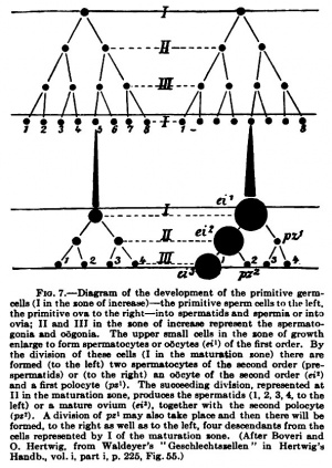
A comparison of the ovium and spermium is possible only when the development of both cells is considered. This will be done in the chapter on the urogenital apparatus and it will be only briefly treated here. In the development of the male and female germ-cells three periods may be distinguished. The first period is that of increase, in which the germ-cells — at this stage termed oogonia in the female and spermatogonia in the male — undergo a rapid increase by mitotic division. Af a certain period of development tlie divisions cease and the cells produced by the last divisions — the oocytes of the first order in the female and the spermatocytes of the first order in the male — then enter upon a period of growth, which, especially in the female, leads to a great increase in size. At the close of this period that of maturation begins, during which the polocytes are formed in the female cell. A corresponding period oCours in spermatogenesis. Two divisions follow quickly on one another; each spermatocyte of the first order divides first into two spermatocytes of the second order and each of these again divides into two spermatids. Just as in the case of the matured ovum, the oide or ovium, and the three polocytes, which are to be regarded as rudimentary oides, the chromatin elements are reduced to half their original number, so too in each of the four spermatids that are formed by the division of a spermatocyte of the first order the chromosomes are reduced by one-half. A marked difference, however, oCours: whereas the four oides (the ovium and the three polocytes) are very unequal in size, the ovium being many times larger than each of the polocytes, the four spermatids from each spermatocyte of the first order are equal in size. And another difference lies in this, that while the ovium is capable of being fertilized immediately after or even during the second maturation division, the four spermatids must still undergo a complicated process of development, that has no equivalent in the ovium; they must be transformed into spermia before they are capable of fertilizing. A comparison of spermatogenesis and oogenesis is clearly shown by Boveri's diagram (Fig. 7), taken from Waldeyer (p. 225).
The male and female germ-cells are shown by this comparison to be essentially equivalent, and yet the subordinate differences are of the greatest importance. Both are cells in which the nimaber of the chromosomes has been reduced to one-half by quite comparable processes ; their differences are associated with a division of labor. In the ovum nutriment is stored up for the new being that will be formed; it consequently grows to a considerable size. During the maturation divisions all the nutritive material is retained by one cell; the polocytes receive none of it, the ovium contains it all. On account of the mass of nutritive material the ovium becomes heavy and is not in a condition to move toward a union with the male germcell ; it must await it. In the spermatocytes of the first order there is, in the first place, much less nutritive material stored up, and, in the second place, it is equally distributed among all the four spermatids during the maturation divisions ; and, in order that they may be able to seek out the ovium, each of these four spermatids must be further modified into spermia. While, therefore, from each oocyte of the first order but one ovium capable of being fertilized is produced, from each spermatocyte four spermia arise, and thus there are formed from an equal number of spermatocytes and oocytes of the first order four times as many fertilizing spermia as there are fertilizable oides. And yet this difference, as we shall see later, does not represent, even approximately, the numerical relations of the mature ova and the spermia.
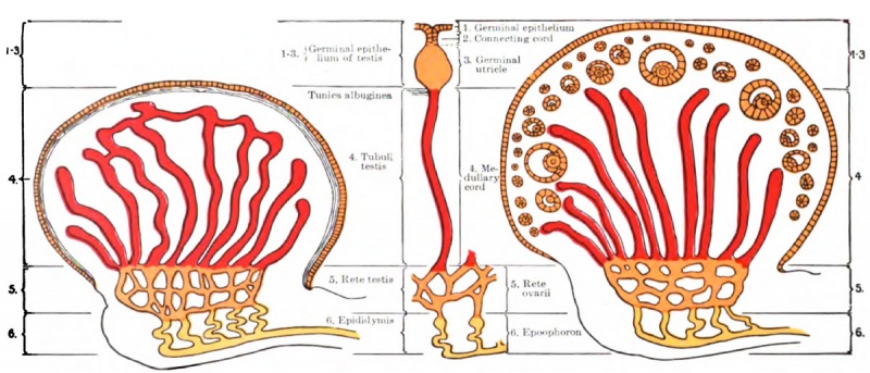
Fig. 8. Diagram showing a comparison of the testis and the ovary (based on the results of Winiwarter and Waldeyer). The germinal epithelium of the texts corresponds to 1-3 of the ovary.
It must be noted, however, that so far as man and the mammals are concerned the comparison of the oogenesis and spermatogenesis shown in Boveri's diagram is not quite free from objection, for it is doubtful if the oogonia and spermatogonia can be exactly homologized in these forms. For although both arise from the germinal epitheliimi, nevertheless they appear to belong to different cell generations. In the male, according to recent observations, the germ-cells have their origin from cells which correspond to those of the medullary cords of the ovary; the ingrowths which give rise to the germinal cords, in which the ova develop, have no homologue in the testis. The annexed diagram will make this clear.
| Online Editor - germinal epithelium |
|---|
|
We now know that the "germinal epithelium" is a misnomer as it is simply the epithelium surrounding the gonad and not the source of germ cells (primordial germ cells) that will form oocyte and spermatozoa. These cells originate elsewhere, probably the first cells to migrate through the primitive streak during gastrulation, lie adjacent to the hind-gut and migrate into the region of the early developing gonad.
|
Bonnet[24] imagines that exceptionally the polocytes may also be fertilized and give rise to embryomata.
As regards the number of germ-cells which may normally be produced, Hensen estimates that a human female develops in each ovary about 200 ova ripe for fertilization. Lode[25] found in 1 c.mm. of a human ejaculate 60,876 spermia, and from that calculates for the entire ejaculate, which averages 3373 c.mm., over 200 million spermia, so that during his life a man may produce about 340 billion spermia* Consequently for every mature ovium there are about 850 million spermia.
Finally, the question is to be considered whether the sex of the future individual is in any way determined in the germ-cells, the ovium or spermium. In the chapter on the development of the urogenital organs it will be shown that with our present methods of observation the sex of the human embryo cannot be determined before the fifth or sixth week of development; as is well known it has been supposed that during the earlier stages of pregnancy influences may be brought to bear which will determine the formation of the one or the other sex. This, however, seems to be impossible, since whether the developing organism shall be of the male or the female sex is, apparently, already determined at fertilization. This is indicated by the fact that twins and double monsters formed from a single ovum are always of the same sex. Most observers now incline to the belief that the sex is always determined before fertilization in the ovium, that there are mature ova (ovia) from which only females and others from which only males can be formed. In my opinion the question is not yet settled so far as man and the vertebrates are concerned, but it is certain that in many invertebrates the sex is already determined in the egg. How liie germ-cells of the human female or male may be influenced so that they will produce either the one or the other sex has been frequently discussed, but none of the conclusions will stand serious criticism. Nussbaum[26] has shown that in the rotifer Hydatina senta the sex of the progeny is determined while they are still within the body of the mother, since all poorly nourished females deposit eggs from which males develop, while from the eggs deposited by well-nourished females only females were formed. All the eggs of any one female produce the same sex and neither fertilization nor any treatment after fertilization has any effect. In an extensive series of observations on a mammal, the mouse, 0. Schultze[27] did not suCoeed in influencing the sex character ; no influence was exerted by the age or the difference of age of the parents, by the age of the sexual products, by in-breeding or by incest-breeding, by sexual appetency, etc. For further consideration of the question reference may be made to Henneberg[28] and Lenhossek.[29]
References
- ↑ W. Nagel: Das menschliche Ei, Arch. f. mikr. Anat., voL xxxi, 1888.
- ↑ Korschelt and Heider : Lehrbuch der vergleichenden Entwicklungsgeschichte, 1902.
- ↑ Haecker: Die Chromosomen als angenommene Vererbungstrager, Ergebnisse und Fortschritte der Zoologie, herausgegeben von J. W. Sprengel, vol. i, 1907.
- ↑ Waldeyer: Chapter I in 0. Hertwig's Handbuch der vergleichenden und experimentellen Entwicklungslehre, 1906.
- ↑ E. B. Wilson : The Cell in Development and Inheritance, New York, 1906.
- ↑ J. Sobotta: Die Bildung der Riehtungskorper im Ei der Maus, Anatom. Hefte, evi, 1907. The remaining literature is cited here.
- ↑ L. Gerlach : Ueber die Bildung der Riehtungskorper bei Mus musculus, Wiesbaden, 1906.
- ↑ Retzius: Zur Kenntnis vom Bau des Eierstockeies und des Graaf'schen Follikels, Hygiea Festband, Stockholm, 1889.
- ↑ Flemming: Zellsubstanz, Kem und Zellteilimg, Leipzig, 1882, p. 35.
- ↑ Von Ebner: Ueber das Verhalten der zona pellucida ziun Ei, Anat. Anz., vol. xviii, 1900.
- ↑ Paladino: La renovazione del parenchima ovarico nella donna, Atti delF XI Congr. internaz. med. di Roma, vol. ii, Anatomia, 1894, p. 19. Compare also Arch. ital. de Biologic, vol. xxi, p. 15, 1894; and Monitore zoolog. ital.. Anno V, p. 140, 1894; also, Per il tipo di struttura delP ovaja, Rendic. Acad. Sc. fis. math., Napoli, vol. iii, p. 232, lvS97; also, Sur le type de structure de I'ovaire, Arch. ital. de Biol., vol. xxix, p. 139, 1898.
- ↑ Bonnet: Gibt es bei Wirbeltieren Parthenogenesis? Ergebnisse d. Anatomie und Entwicklungsgeschichte, vol. ix, 1900.
- ↑ See, also, Meves : Zur Entstehung der Achsenf aden menschlicher Spemiatozoen, Anat. Anz., vol. xiv, 1897 ; and Ueber das Veriialten der Centralkorper bei der Histog«nese der Samenfaden von Mensch und Ratte, Verb. Anat. Ges. (Kiel), 1898.
- ↑ J. Duesberg: Sur le nombre des chromosomes chez Phomme, Anat. Anz., vol. xxviii, 1906.
- ↑ E. M. Nelson: Some Observations on the Human Spermatozoon, Joum. Quekett Micr. Club, London, ser. 2, iii, pp. 310-314, 1889.
- ↑ Von Bardeleben: Ueber die Entstehung der Achsenfaden im menschlichen und Saugetierspermatozoon, Anat. Anz., vol. xiv, 1897; also, Beitra^ zur Histologic des Hodens und zur Spermatogenese beim Menschen, Arch, f . Anat. und Entwicklungsgesch., Supplementband, 1897; also, Weitere Beitrage zur Spermatogenese beim Menschen, Jenaische Zeitschrift, vol. xxxi, 1898.
- ↑ Gustav von Widersperg: Beitrage zur EntwicklungsgeEchichfo der Samenkorper, Areh. f. mikr. Anat., vol. xxv, 1885 (PI. VI, Fig. 18).
- ↑ G. Retzius: Zur Kenntnis der Spermatozoon, Biol. TJntersueh., 1881.
- ↑ Ivar Broman: Ueber atypiscbe Spermien (speziell beim Menschen) und ihre mbglirhe BedeutuDg, mit 107 Abbildungen, Anat. Anz., vol. xzi, p. 455—161, 1902.
- ↑ Von Bardeleben: Dimorphism us der mannlichen Oeschlechtszellen bei Saugetieren, Anat. Anz., vol. ziii. 1897.
- ↑ A. Roth : Ueber das Verhalten beweglicher Mikroorganismen in stromenden Flussigkeiten, Deutsche med. Wochenschrift, Jg. 19, p. 351-352, 1893; also, Zur Kenntnis der Bewegung der Spermien, Arch. f. Anat. und Physiol., Physiol. Abt., 1904.
- ↑ H. Adolphi : Die Spermatozoen der Saugetiere schwimmen gegen den Strom, Anat. Anz., vol. xxvi, 1905.
- ↑ Bossi : fitude clinique et experimentale de Fepoque la plus favorable h la fecondation et de la vitalite des spermatozoi'des sejoumant dans le nidus seminis, Rivista di ostetric. e ginecoL, 1891, no. 10, also in Nouv. Arch, d'obstetr. et de gynecoL, April, 1891.
- ↑ Bonnet: Zur Aetiologie der Embryome, Greifswald. med. Verein, Bericht in Miinchener Med. Wochenschrift, 1901, p. 315.
- ↑ A. Lode: Untersuehung:en Uber Zahlen und R^renerationsverhaltnisse der Spermatozoiden bei Hunde und Mensch, Arch. f. jjes. Phys., 1891; also, Experinientelle Beitrage zur Physiologie der Samenblasen, Sb. K. Acad. Wiss. Wien., Abt. 3, vol. civ, p. 33, 1895.
- ↑ M. Nussbaum : Die Entstehung des Geschlechts bei Hydatina senta, Arch, f . mikr. Anat., vol. xlix, p. 227-308, 1897.
- ↑ 0. Schultze : Zur Frage von den geschlechtsbildenden Ursachen, Arch, f . mikr. Anat., vol. Ixiii, p. 197-257, 1903.
- ↑ B. Henneberg: Wodurch wird das Geschlechtsverhaltnis beim Menschen und bei den hoheren Tieren beeinflusst? Ergebnisse der Anat. und Entwicklungsgesch., vol. vii, p. 697-721 (Lit., 1897), 1898.
- ↑ M. von Lenhossek: Des Problem der geschlechtsbestimmenden Ursachen, Jena, 1903.
| Historic Disclaimer - information about historic embryology pages |
|---|
| Pages where the terms "Historic" (textbooks, papers, people, recommendations) appear on this site, and sections within pages where this disclaimer appears, indicate that the content and scientific understanding are specific to the time of publication. This means that while some scientific descriptions are still accurate, the terminology and interpretation of the developmental mechanisms reflect the understanding at the time of original publication and those of the preceding periods, these terms, interpretations and recommendations may not reflect our current scientific understanding. (More? Embryology History | Historic Embryology Papers) |
Glossary Links
- Glossary: A | B | C | D | E | F | G | H | I | J | K | L | M | N | O | P | Q | R | S | T | U | V | W | X | Y | Z | Numbers | Symbols | Term Link
Cite this page: Hill, M.A. (2024, April 27) Embryology Book - Manual of Human Embryology 1. Retrieved from https://embryology.med.unsw.edu.au/embryology/index.php/Book_-_Manual_of_Human_Embryology_1
- © Dr Mark Hill 2024, UNSW Embryology ISBN: 978 0 7334 2609 4 - UNSW CRICOS Provider Code No. 00098G

