Book - Human Embryology and Morphology 2
| Embryology - 27 Apr 2024 |
|---|
| Google Translate - select your language from the list shown below (this will open a new external page) |
|
العربية | català | 中文 | 中國傳統的 | français | Deutsche | עִברִית | हिंदी | bahasa Indonesia | italiano | 日本語 | 한국어 | မြန်မာ | Pilipino | Polskie | português | ਪੰਜਾਬੀ ਦੇ | Română | русский | Español | Swahili | Svensk | ไทย | Türkçe | اردو | ייִדיש | Tiếng Việt These external translations are automated and may not be accurate. (More? About Translations) |
Keith A. Human Embryology and Morphology. (1902) London: Edward Arnold.
| Historic Disclaimer - information about historic embryology pages |
|---|
 |
| Online Editor |
|---|
|
Links: Respiratory System Development
Chapter II. The Nasal Cavities and Olfactory Structures

In tracing the development of structures subservient to the sense of smell, the following elements have to be dealt with :
- The olfactory sense epithelium and olfactory nerves ;
- The parts of the brain concerned with the sense of smell, so far as we know them ;
- The capsule which contains the olfactory epithelium ;
- The respiratory tract of the nasal cavities.
Origin of the Olfactory Sense Epithelium
A small area of the epiblastic cells, lying under the fore-brain becomes demarcated on each side, to form the olfactory plates. Around these two plates the lateral and mesial nasal processes grow up (Fig. 16), the plates being depressed to form the olfactory pits. With the growth of the nasal processes the olfactory plates and pits are thrust into the roof of the stomodaeum, where they form the epithelial lining of the upper third or olfactory area of the nasal cavities. A small island is detached from each plate to form the basis of Jacobson's organ (Fig. 16). The sense epithelia send out nerve processes which form arborescences round the neural cells of the outgrowing olfactory lobe (Fig. 17). The olfactory nerves are thus formed. At first the olfactory plate is directly in contact with the neural tube, and it is probable that neuroblasts may migrate then to the olfactory plate and form the olfactory sense epithelium.
The condition of olfactory pits, a developmental phase in the human embryo, is the permanent form in fishes (Fig. 21 B) ; in amphibians and all higher vertebrates the fundus of the pit breaks down and thus the olfactory pits come into communication through the posterior nares with the stomodaeum (Fig. 8).
The Olfactory Lobe
As the olfactory plates are being thrust downwards, the anterior part of the floor of the fore-brain grows out on each side as a hollow protrusion to form the olfactory lobes (Fig. 145). At the end of the 3rd month the olfactory lobe has assumed the form shown in Fig. 17. Its cavity is at first continuous with that of the cerebral vesicle (lateral ventricle), but this connection is soon lost; it becomes solid and divided into anterior and posterior parts by a transverse fissure (Fig. 17).

Fig. 17. The Mesial aspect of the Brain of a human foetus, 3.5 months old, showing the Olfactory Lobe.
The anterior part, as is shown in figure 18, becomes (a) the olfactory bulb, (b) the olfactory peduncle or tract, (c) the trigonum olfactorium, lying between the lateral and mesial roots into which the tract divides, and (d) the area of Broca. The posterior part of the olfactory lobe (B in Fig. 17) becomes (a) the grey matter of the anterior perforated space, and (b) the gyrus subcallosus or peduncles of the corpus callosum (Fig. 18).
Termination of the Olfactory Tract
As is shown in figure 18, the mesial root terminates in the supra-callosal gyrus and fornix, while the lateral ends in the uncus of the hippocampal convolution. Olfactory nerve fibres also terminate in the trigone and area of Broca. To the parts derived from the olfactory lobe together with the uncus, the fascia dentata, the supra-callosal gyrus, the fimbria, the fornix and septum lucidum (Figs. 18 and 172) the term Rhinencephalon is given because these" parts are concerned with the sense of smell, and represent the parts first and most highly developed in the brains of lower vertebrates (Elliot Smith). In man many of these parts are merely vestigial. The higher olfactory centre has been located in the hippocampal gyrus, but no evidence has been given showing any connection between the callosal gyrus and this sense. In animals with a highly developed olfactory sense (carnivora, etc.) these parts of the brain which form the rhinencephalon are well developed. The fornix in its greater part, and the longitudinal striae as association tracts, connect the brain areas which are subservient to the sense of smell (see Fig. 18).

Fig. 18. Showing the parts formed out of the Olfactory Lobe in the brain of an Adult, and the termination of the olfactory roots in the Sub-callosal and Uncinate Gyri. (After Elliot Smith)
It is important, from a clinical point of view, to remember that the olfactory nerves are surrounded by prolongations of the arachnoid membrane and subarachnoid spaces, and through these spaces infections may spread from the nasal cavities to the meninges. Further, the olfactory tracts rest on the edges of the small wings of the sphenoid, and may be injured in falls on the forehead..
The Nasal Cavities
The separation of the nasal cavities from the stomodaeum by the downgrowth of the mesial and lateral nasal processes, and the ingrowth of the horizontal plates of the maxillary processes, has been already described (p. 3). These processes also form and bound the anterior and posterior nares.
Development of the Nasal Air Sinuses
The manner in which the nasal mucous membrane pushes its way through the lateral nasal cartilage into the maxillary process to form the Antrum of Highmore has been already described (page 12). The other air sinuses — the frontal, lachrymo-ethmoidal, anterior, middle and posterior ethmoidal, and sphenoidal sinuses — six in all, arise in the same way as the antrum but at a much later date. Although they begin to bud out about the 3rd year, they assume their active growth in the earlier years of puberty, and reach their full size before the 30 th year.
At birth, the lateral mass of the ethmoid is a thin plate, carrying the superior and middle turbinate processes, which almost fill the nasal cavity (Fig. 7). The entire ethmoid is narrow, and hence the proximity of the eyes in children. Beneath the middle turbinate is a thumbnail-like impression — the hiatus semilunaris (Fig. 19). The antrum buds out near its posterior end, arid the point at which the bud starts becomes its opening. The uncinate process of the lateral mass of the ethmoid forms the prominent lower margin of the hiatus.
From the upper end of the hiatus a bud of mucous membrane grows upwards to form the frontal sinus, gradually works through the ethmoid, and pushes its way into the frontal bone, separating the outer from the inner lamellae. As a rule, by the 25 th year it reaches outwards over the inner two-thirds of the orbital roof, and is an inch or more both in height and depth at its inner part. It is smaller in women than in men, but it may be, and often is, arrested at an early stage of development, or it may be absent altogether. The size of the glabellar prominence is no index to its development.

Fig. 19. A diagram of the Lateral Wall of the Nasal Cavity, showing the position of the Air Sinus. The parts beneath the turbinate processes are indicated by stippled lines.
The stalk of the frontal bud forms the infundibulum, which is narrow, half an inch long, and difficult of catheterization from the nose. Into it open (or sometimes into the hiatus) the lachrymoethmoidal and anterior ethmoidal cells which surround the infundibulum. They are developed as outgrowths from the infundibulum (Fig. 19). Occasionally the antrum of Highmore, as is frequently .the case in the gorilla, sends a process to form part of the frontal sinus, and hence there may be a communication between the sinus and the antrum.
The development of the frontal sinuses and supra-orbital ridges lead to a marked change in the face at puberty. By the formation of the frontal sinuses the basal area of the skull, to which the face is attached, is largely increased in extent. Such an increase is necessary to support the palate, which grows rapidly in size at puberty. Up to the fifth year the upper jaw has to carry only ten milk teeth ; in the adult it has to carry sixteen permanent teeth. To support these the face and palate grow rapidly in size. The formation of the frontal sinus gives the necessary increase in the area of the base of the skull for their support. It should be remembered that the growth of the brain and of the cranial cavity is comparatively slight after the fifth year.
Only the gorilla and chimpanzee show an arrangement of frontal and ethmoidal sinuses comparable to that of man.
Above the hiatus lies the bulla ethmoidalis, which is inflated by, and commonly carries the opening of, the middle ethmoidal cell (Fig. 19). The posterior ethmoidal sinus opens beneath the superior turbinate process, and is developed from the superior meatus. The ethmoidal sinuses are produced in the cartilage of the ethmoidal or lateral nasal plate (Fig. 7). They inflate the ossifying cartilaginous plate until it becomes a cellular mass, thus increasing the breadth of the intra-orbital septum. The .sphenoidal sinus (Fig. 19) is formed by the mucous membrane growing into and expanding the sphenoidal turbinate bone, which is a small, slightly ossified cartilage lying beneath the presphenoid at birth, and forming the uppermost (sixth) of the nasal turbinate processes. Latterly the sinus grows into and expands the pre-sphenoid and part of the basi-sphenoid, the sphenoidal turbinate remaining as its anterior wall (Fig 19). The •sphenoidal turbinate is a detached part of the ethmoidal (lateral nasal) cartilage.
It will thus be seen that all the nasal air sinuses are produced primarily by a budding outwards of the nasal mucous membrane into the cartilaginous basis of the lateral nasal processes. Disease may readily spread to these sinuses from the nasal cavities. By means of the sinuses the area of the face is increased to support the adult palate bearing the permanent teeth. Most of them open on the respiratory tract of the nasal cavity. They are ventilated with every breath. They act also as resonance chambers.
Vestigial Turbinates
There is frequently to be seen in the adult one, or even two, secondary meatuses above the superior ; these are constantly present in the chimpanzee and in animals with a keen sense of smell. In the human foetus of four months six turbinates are usually present, besides secondary processes in the meatuses beneath them. The uppermost of these, the sixth, becomes the sphenoidal turbinate ; the fifth disappears ; the third and fourth may remain separate or become united ; the first and second form the inferior and middle turbinate processes. The agger nasi (naso-turbinal, Fig. 19), in front of the attachment of the middle turbinate process, is a vestige of the naso-turbinal, a process well developed in most carnivora and animals with a strong scent. The uncinate process, which forms the lower border of the hiatus semilunaris, is continuous at its base with the naso-turbinal. Through the hiatus semilunaris acting as a gutter, the antrum may become a cesspool for a suppurating frontal sinus.
Nasal Duct
Although in no way connected with the sense of smell, the nasal duct is closely related to the nasal cavities.

Fig. 20. Showing on the inner wall of the Orbit (1) the position of the Infundibulum, (2) the pars facialis lachrymalis.
It is formed between the lateral nasal and maxillary processes (Figs. 1 and 7). Three bones bound it : the superior maxilla on the outer side, formed in the maxillary process; the inferior turbinate, formed in the cartilage of the lateral nasal process, and the lachrymal, formed over the lateral nasal cartilage, bound it on the inner side. The formation of the palate cuts the duct off from the mouth. The hamulus of the lachrymal varies much in size, and is the vestige of a larger process, which in lower primates enters into the formation of the inferior margin of the orbit. This pars facialis sometimes occurs in man (Fig. 20). The position of the infundibulum to the lachrymal is shown in Fig. 2 ; it is seen to lie entirely in the lateral mass of the ethmoid behind the lachrymal. Occasionally the frontal and superior maxillary bones may articulate in an interval between the lachrymal in front and lateral mass of the ethmoid behind.
Chapter Figures
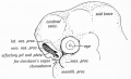
Fig. 16. The Olfactory Pit and Nasal Processes in a 4th week human embryo.
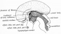
Fig. 17. The Mesial aspect of the Brain of a human foetus, 3J months old, showing the Olfactory Lobe.
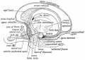
Fig. 18. Showing the parts formed out of the Olfactory Lobe in the brain of an Adult, and the termination of the olfactory roots in the Sub-callosal and Uncinate Gyri.
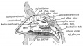
Fig. 19. A diagram of the Lateral Wall of the Nasal Cavity, showing the position of the Air Sinus.
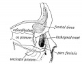
Fig. 20. Showing on the inner wall of the Orbit (1) the position of the Infundibulum, (2) the pars facialis lachrymalis.
- Links: Smell Development
| Historic Disclaimer - information about historic embryology pages |
|---|
|
Human Embryology and Morphology (1902): Development or the Face | The Nasal Cavities and Olfactory Structures | Development of the Pharynx and Neck | Development of the Organ of Hearing | Development and Morphology of the Teeth | The Skin and its Appendages | The Development of the Ovum of the Foetus from the Ovum of the Mother | The Manner in which a Connection is Established between the Foetus and Uterus | The Uro-genital System | Formation of the Pubo-femoral Region, Pelvic Floor and Fascia | The Spinal Column and Back | The Segmentation of the Body | The Cranium | Development of the Structures concerned in the Sense of Sight | The Brain and Spinal Cord | Development of the Circulatory System | The Respiratory System | The Organs of Digestion | The Body Wall, Ribs, and Sternum | The Limbs | Figures | Embryology History
Reference
Keith A. Human Embryology and Morphology. (1902) London: Edward Arnold.
Cite this page: Hill, M.A. (2024, April 27) Embryology Book - Human Embryology and Morphology 2. Retrieved from https://embryology.med.unsw.edu.au/embryology/index.php/Book_-_Human_Embryology_and_Morphology_2
- © Dr Mark Hill 2024, UNSW Embryology ISBN: 978 0 7334 2609 4 - UNSW CRICOS Provider Code No. 00098G