Book - Contributions to Embryology Carnegie Institution No.22
| Embryology - 2 May 2024 |
|---|
| Google Translate - select your language from the list shown below (this will open a new external page) |
|
العربية | català | 中文 | 中國傳統的 | français | Deutsche | עִברִית | हिंदी | bahasa Indonesia | italiano | 日本語 | 한국어 | မြန်မာ | Pilipino | Polskie | português | ਪੰਜਾਬੀ ਦੇ | Română | русский | Español | Swahili | Svensk | ไทย | Türkçe | اردو | ייִדיש | Tiếng Việt These external translations are automated and may not be accurate. (More? About Translations) |
Wheeler T. Study of a human spina bifida monster with encephaloceles and other abnormalities. (1918) Contrib. Embryol., Carnegie Inst. Wash., 22: .
| Online Editor |
|---|
| This historic 1918 paper by Wheeler describes an example of human embryo spina bifida. This was an early publication in the series from the Carnegie Institution of Washington called Contributions to Embryology. The term "monster" is the historic term for gross developmental abnormalities and is no longer used and is also offensive. It has been left within this text as this is how such major abnormalities were medically referred at that time, and I apologise for any offence to the reader that use of this term may cause.
|
| Historic Disclaimer - information about historic embryology pages |
|---|
| Pages where the terms "Historic" (textbooks, papers, people, recommendations) appear on this site, and sections within pages where this disclaimer appears, indicate that the content and scientific understanding are specific to the time of publication. This means that while some scientific descriptions are still accurate, the terminology and interpretation of the developmental mechanisms reflect the understanding at the time of original publication and those of the preceding periods, these terms, interpretations and recommendations may not reflect our current scientific understanding. (More? Embryology History | Historic Embryology Papers) |
Study of a Human Spina Bifida Monster with Encephaloceles and Other Abnormalities
Carnegie Institution of Washington - Contributions to Embryology
By Theodora Wheeler. With four plates
--Mark Hill 01:02, 16 February 2011 (EST) Please note the term "monster" is the historic term for gross developmental abnormalities and is no longer used and is also offensive. It has been left within this text as this is how such major abnormalities were medically referred at that time, and I apologise for any offence to the reader that use of this term may cause.
Introduction
The specimen described in this study is a human female monster with spina bifida, in which there is total subcutaneous involvement of the spine and a defective occiput. The thoracic and cervical regions of the spine are much shortened, and encephaloceles and numerous other abnormalities are present. The type, though a rather unusual variety of spina bifida, occurs frequently enough to have been recognized and grouped by itself for some time past, and to this group the term iniencephaly has been applied. Because of the striking appearance of these specimens, one or more are usually to be found in any museum of pathology. In the embryological collection of over 1,600 specimens belonging to the Department of Embryology of the Carnegie Institution of Washington, the only example is the one presented in this paper. No. 862a. It was through the courtesy of the Bridgeport General Hospital that this specimen was obtained.
Only a short review of the literature on spina bifida will be given here. More complete historical accounts with extensive bibhographies are to be found in articles by Kermauner and by Ernst in Schwalbe's IMorphologie der Missbildung (1909), and also in a chapter on spina bifida by Tillmann, in his volume of Deutsche Chirurgie, v. 62a (1905). The earliest references to many teratological conditions are supposed to be found in folklore and in mythological tales of centaurs, Cyclops, mermaids, and such creatures, and it has been suggested that among such stories may likewise be found the first record of spina bifida. Possible the hairy and cloven-hoofed satyr was originally a fairly normal individual with spina bifida, hypertrichosis, and club feet, whose abnormalities gradually developed through excited hearsay into the hind-quarters of a beast. Even in recent times, in connection with scientific work on this condition, such inaccuracies have only too frequently been paralleled by superficial observations and indefinite speculations. However, it is not surprising that a good deal of vagueness has existed in regard to spina bifida, as the subject-matter includes widely dissimilar and very comphcated conditions.
As described among human forms, two chief types are distinguished : the flatspine type (rachischisis, spina bifida aperta) and the subcutaneous type (cystic, occulta). In both of these forms the greatest variations exist as to location and amount of spinal involvement. In some instances only a single segment is affected ; in others the whole spinal column, together with the cranium, may be involved. Combinations of these two forms are to be found, and also conditions where the two varieties merge into one another. Associated with every type of the condition are found innumerable other abnormalities.
Owing to this mass of complicated material and to the widely different nomenclatures used by the large number of investigators who have worked on the problem,
the literature is enormous and rather confused. The classification is still very superficial. In teratology, as in general pathology, the trend has been to supplant classifications based on regional distribution by those having an etiological basis. There exists still in the literature on spina bifida a great deal of the former method. This is due to the fact that until quite recently study has been of external form alone, from which method only a crude regional classification can result. By the application of the more penetrating methods of modern anatomy, embryology, and experimental biology, progress has been made toward etiological classification.
In 1881 Koch assembled a number of different forms of spina bifida. He pointed out the distinction between the flat-spine form, in which the spinal cord is uncovered (spina bifida aperta), and the cystic or subcutaneous form, in which the soft parts have joined but the bony arches remain ununited. He attributed a later formative period to the subcutaneous than to the open form. In 188G von Recklinhausen presented over 30 specimens of spina bifida and focussed attention especially upon the pathological anatomy of the central nervous system and its membranes in the fetal and older forms. By thoroughly analyzing the conditions met with and applying the conception of arrested development, he was able to offer reasonable interpretations for much of the developmental mechanism which up to that time had not been understood. Contemporaneously with these two writers, and since their time, many aspects of the subject have been studied. The surgical treatment of spina bifida has been taken up by many, notably Bayer, Hildebrand. and Muscatello. Other authors have described special types of the abnormality Among these may be mentioned Lewis's paper on iniencephaly. He collected 23 cases similar to the one herein described, which show some of the variations presented by this special form. In the literature are to be found fairly numerous descriptions of young specimens with spina bifida. In "A study of the causes
underlying the origin of human monsters" (1908), Mall describes 12 from his collection and cites several others from the literature. An interesting 8 mm. ferret embryo with localized cervical hydromyelia was described by Good in 1912.
Experimental studies on the lower animals have formed a very important source of information with regard to the open variety of spina bifida. In Mall's earlier a review of the literature on the subject up to 1908 is given. The work of Hertwig and Morgan has attracted especial attention, the former showing that external agents causing delay in the closure of the blastopore can bring about embryological spina bifida. The work of the latter author has been interpreted as pointing toward NaCl as the definite etiological agent, as he was able to produce a delayed closure of the blastopore in frogs' eggs through the use of a 0.6 per cent solution of XaCl. Embryological spina bifida has also been produced occasionally in chicks by overheating and various other methods. Working with frogs' eggs, by ultra-violet-ray exposures Baldwin (1915) obtained a condition of doubled and closed neural canal aiul sometimes doubled cord. His specimens were usually two-tailed. He referred to them a.s spina bifida and gave a clear explanation of the mechanics of the process producing- them. However, the relation of this type of spina bifida to the more ordinary condition of a single open neural canal is not altogether plain, and his suggestion that " imperfect oxidation " causes spina bifida does not further claiify the question.
The earliest picture of the subcutaneous type of spina bifida with which we are familiar is that encountered in embiyos around the 10 mm. stage of development, in which the neural tube is everywhere closed, showing, however, a greater or less area of enlargement. Such a state has not as yet been experimentally produced. Several explanations have been advanced to account for it, none of which are satisfying, nor substantiated by evidence. One suggestion is that the enlargement of the neural tube is due to the fact that dorsally it remains attached to the ectoderm (non-separation of the membrana reuniens). On the other hand, it has been suggested that the neural tube becomes enlarged because of increased pressure from the contained fluid. In this connection it would seem that when the affected areas are limited in extent they are in some way connected with the curvatures of the body, since such areas usually occur in the neck or sacral region, where, in the embryo, the curves are most pronounced. The process is supposed to be one of subsequent pressure of the h3alromyelia on the surrounding parts, thus inhibiting the development of cartilage and bone. With our advance in knowledge regarding- the circulation of the cerebro-spinal fluid, some of the most puzzling features presented by subcutaneous spina bifida will probably be satisfactorily exjilained. The work of Weed on the normal cerebro-spinal fluid circulation is most helpful, supplying as it does for the first time an adequately correlated picture of the formation and extension of the cerebro-spinal fluid with the differentiation of the perimedullary mesenchyme to form the meninges. In the meantime, an}' discussion concerning the etiology of subcutaneous spina bifida is entirely theoretical. Suggestions have been made that it may arise directly from the open spina bifida form; again, that it may be the result of some entirely different process, or that both forms may-be prodiiced by the same pathological agent acting at a different stage of development. While it has been generally assumed that the open and subcutaneous forms of spina bifida are related, this has never been proved. A more definite picture of each process must be obtained before we can know the nature of the relation, or whether there is such a relation, existing between the two. The hterature on the subject gives the impression that, although sound facts and more or less sound theories regarding spina bifida have multiplied, there is much that is not clear and that must be understood before we can have a comprehensive insight into the processes producing it. That this information may be gained through a closer embryological study seems probable.
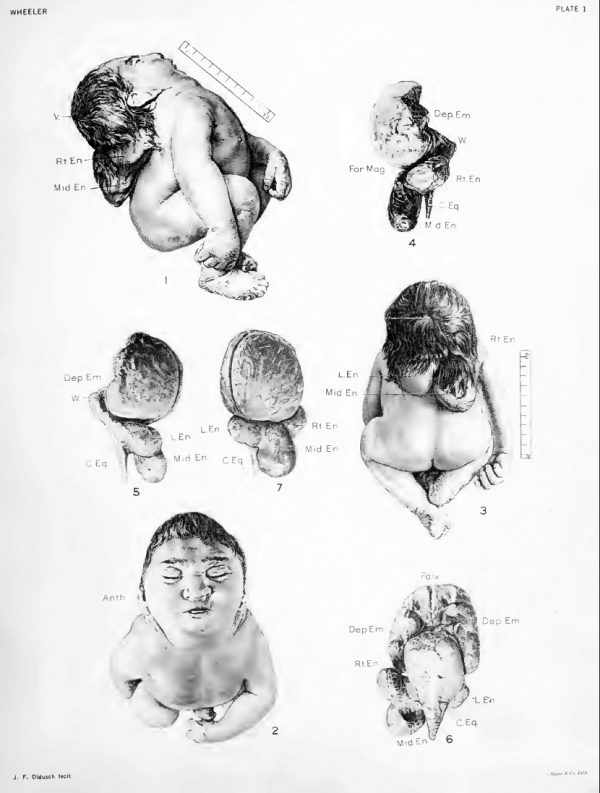
The study of the specimen dealt with in this paper has been made chiefly along morphological lines. Only a meager clinical history regarding it was obtainable; the child was illegitimate, was born spontaneously at full term, and lived only a few hours. Its external form is shown in figures 1, 2, and 3, and various measurements are given in table 2. Externally, the most marked abnormality is the extreme dorsal flexion and shortening of the trunk. The head is drawn back close to the sacral region. The chest and abdomen are unusually prominent. The arms and legs are symmetrical and well developed, but the shoulders are hunched up and lie far forward, close to the cheeks. The face is directed upward, which throws the top of the head back so that the vertex lies level with the raised shoulders. The neck is obhterated and the chin and chest lie in one plane. The features are well formed with the exception of lack of prominence of the chin and deformity of the ears. Figures 1 and 8 show the right ear. The deformity of the left ear is similar. The anthelix is pushed outward so as to be unusually prominent; the tragus is shifted medially and upward, so that it lies opposite the concha; the antitragus lies below it, pressed against the cheek. Darwin's tubercle is present. The external auditory meatus is patent and the parts of the middle and inner ear prove on dissection to be well developed. The whole external ear is considerably narrowed, as is indicated by as low a physiognomical index as 48.5. The average physiognomical index of the right ear of 14 white infants under 3 weeks of age, in the obstetrical ward of the Johns Hopkins Hospital, was found to be 69.1, varying between 62.5 and 78.7. Measurements made by Dr. A. H. Schultz of 4 dead white infants not older than 1 month showed the physiognomical index of the right ear to be 65.0, with a variation between 60.0 and 73.1. Though the physiognomical index shows a rather wide variation due to the great flexibility of the ear cartilage in infants, nowhere in the small group of available normal cases is it nearly so low as in his specimen. The ear deformity is apparently caused by pressure upon and twisting of the external parts of the ear during their development by the backward-bent head and the shoulders which lie close on either side. Marx describes a deformed ear which he designates as "Wildermiithsche Ohr, " in which the anthelix is very prominent. From the base of each ear a crease in the skin extends for 3 cm. medially under the chin to within 2 cm. of the midline, as is seen in figure 2.
Content to be added----
Explanation of Plates
Plate 1
Fig. 1. Right lateral view of specimen shows extreme dorsal flex-ion with vertex level with shoulders Middle and right encephaloceles show in this view. The distorted right ear here seen is drawn in detail in figure 8.
Fig. 2. Superior view of the specimen looks directly at the face Measurements given in table 1 . Transversely across the forehead at the hair line an artefact puckering e.xtends horizontally for 2 cm.
Fig. 3. The dorsal view shows the shortened trunk, superior surface of head, and encephaloceles
Fig. 4. Right lateral surface of subdural cast, showing middle and right encephalocele
Fig. 5. Left lateral surface of subdural cast, showing middle and left encephalocele
Fig. 6. Ventral surface of subdural cast
Fig. 7. Dorsal view of subdural cast. The falx is seen to be to the left of midline
Original plate images were at half actual size magnification.
Plate 2
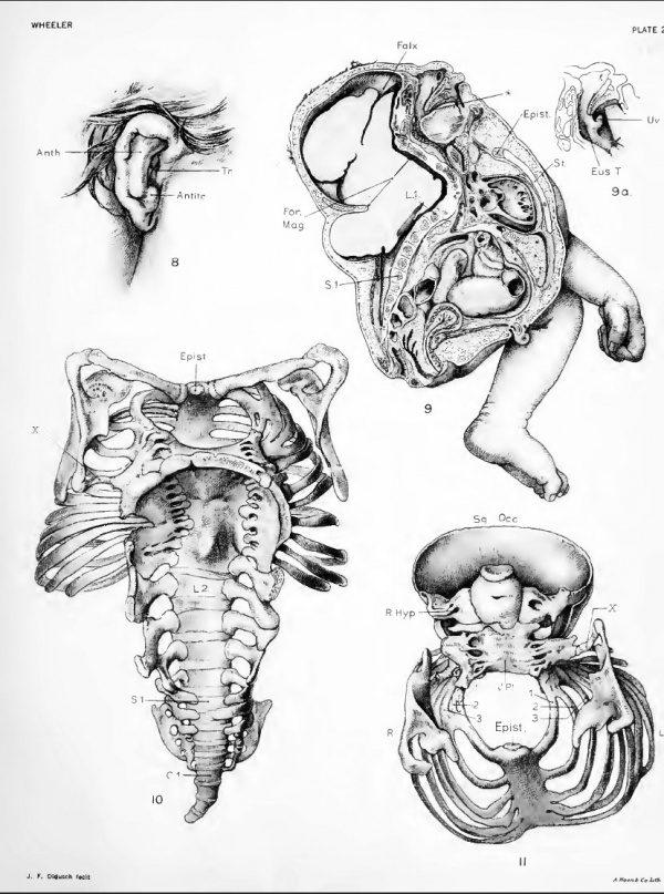
Fig. 8. Sketch of right ear (natural size), showing the anthelix unusually prominent. The tragus lies relatively higher than normal, over rather than horizontally opposite the antitragus. The whole ear very narrow.
Fig. 9. Sagittal section Main outlines were geometrically projected and detail drawn free-hand. The viscera
retain approximately their normal position, absence of the soft palate is shown. The tip of the tongue
lies over the left anlage of the spht uvula. The vertebral column is bent and shortened and irregularly fused
in its upper part. The arches of all the vertebrse are lacking. A fibrous band lies over the upper sacral
vertebra, joining the opposing defective arches in that region and forming a short spinal canal. The section
passes to the left of the sella turcica. The falx cerebri is seen well over on left side. The outhne of the central
nervous system, as is here shown, is used reversed for a diagram in figure 24. The section passes near the
median margin of the left sac. (half original size)
Fig. 9a. Gives left side of bilateral anlage of uvula and orifice of eustachian tube.
(Natural size)
Fig. 10. Shows a dorsal view of the mounted skeleton, with scapulae in place
Varying degrees of gaping vertebral arches are shown at different levels of the spinal column. In the cervical and thoracic regions defective vertebral arches are fused together and markedly everted. In the upper lumbar region they are individually distinct, but still widely everted, while in the lower lumbar and sacral regions they are distinct and bent toward one another. The lumbar transverse processes and the sacral lateral processes are well developed and the coccyx of four segments is seen bent well to the left. In the lower thoracic region a cartilaginous spur projects dorsa'vards from the vertebral bodies. All the thoracic and cervical vertebral bodies are fused together in a single plate. A sUght lateral bending in this plate is present. The foveal surfaces of the atlas face the reader. The intervertebral foramina show large spaces in the lumbar region, which are a sharp contrast to the tiny areas of the contracted thoracic intervertebral foramina. On the right, the rough surface of tho tip of the first lumbar arch is shown, which joins the occiput; and on I lie left, the second lumbar arch, whicli does the same. Crowding of the base of the ribs may be seen, including the first to the sixth on the right and the fifth to the ninth on the left. The sternum is considerably to the left of the milline. A persistent epistermmi is i)resent as a small cartilaginous knob, surrounding the manubrium. The irregular vertebral and superior margins of the scapula; are shown. On the left side the spicule of bone passes from the thoracic and cervical arches to the scapula. (Natural size).
Fig. 11. The superior view of the thoracic skeleton and the anterior surface of the cervical vertebral iilate and of the occiput In the cervical part, no vertebral bodies are distinct, but irregular radicular, and transverse processes project laterally from the central plate. The abnormal spicule of bone on the left side may be seen passing from the fused transverse processes to the left scapula. This view shows how the foveal surfaces of the atlas are shifted to the right in relation to their underlying transverse processes. The right fovea almost overlies the tip of the right transverse process, while the left fovea leaves the left transverse process uncovered. The left transverse process is bent up and joins the pars lateralis, thus forming a rather large foramen. The anterior surface of the occiput shows an asymmetrical oval outline pierced by a foramen, in its center. The double exit of the right hypoglosssal canal shows. The irregular superior margins of the scapula are seen. The episternum and the aborted second rib are demonstrated. (Natural size).
Original plate images magnification shown in brackets.
Plate 3
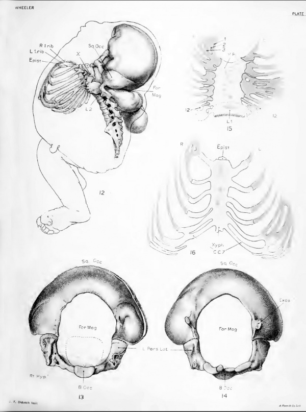
Fig. 12. The left-hand view of the axial skeleton with subdural cast in place and median outline of specimen given. This figure shows the extreme dorsoflexion of the vertebral colunm. The occiput is in position and its squamosal junction on the left with the second lumbar arch is shown. The origin of the spicule of bone which projects out from the transverse process in the thoracic region is visible. The crowding and irregular arrangement of the fifth to the ninth ribs is shown.
Fig. 13. The superior view of the occipital bone shown with its enlarged foramen magnum. A normal-sized foramen
is designated by a dotted line. The left jugular process is prominent when compared with the right, which
seems to have been twisted over to the side. The anterior outlet to the right hypoglossal canal is shown with
the tiny rod of bone which divides the exit of the hypoglossal foramen on the right side immediately under it.
Fig. 14. The inferior surface of the occipital bone shows the large foramen On the squamosal surface the exostoies which join the lumbar vertebra show. On the partes laterales the condylar surfaces and on the left side the cartilaginous process which joins the transverse process of the atlas may be seen. The notched basal margin of the basiocciput also is visible. (Natural size).
Fig. 15. This shows schematically the ventral surface of the thoracic vertebral plate with pairing of the origin of the seventh and eighth and ninth and tenth ribs on the right side, and of the first and second, and the third and fourth on the left.
Fig. 16. This shows schematically the sternum with six costal cartilage attachments on each side The last attachment on both sides is that of the seventh rib. The discrepancy occurs through the second rib becoming aborted on the right side, and the sixth being aborted on the left side. There are four centers of ossification on the midUne of the sternum. The upper two arc opposite the first costal cartilage and resemble an exclamation
mark. The lower two are oval (4 by 3 mm.), with long diameter perpendicular. One is about at the middle
point of the sternum and the other 1 cm. below it an episternum surmounts the sternum and the xyphoid
process projects at its inferior end.
Plate 4
Fig. 17. Dorsal superior view of a normal left scapula of a new-born
Fig. 18. Same view of left scapula of specimen 862a shows the irregular vertebral and superior margins with the
abnormal spicule of bone attached. It also shows the sheets of fascia attached to the vertebral and median
margins of the scapula and the insertions of the rhomboideus and levator scapular muscles on this fascia.
(Natural size).
Fig. 19. Dorsal superior view of a normal right scapula of a new-born
Fig. 20. Same view of right scapula of this specimen showing irregular vertebral margin. (Natural size).
Fig. 21. Diagram of left thoracic and deep dorsal musculature Diagram of left thoracic and deep dorsal musculature on (he left side) of the mounted axial skeleton. The
occiput and model of cerebral-spinal cavity are in place. The median outline of the .specimen is also given in
relation to these structures. Those nuiscles approximately normal arc either sectioned or oidy drawn at
their origin or insertion. They are the m. pectoralis major and minor, the rectus, the external oblique, the
latissimus dorsi, the quadratus lumborum, and the levator scapulae. The abnormal muscles are shown entire,
except for the serratus anterior, whose origin is indicated by broken lines. The largest mass of abnormal
muscles consists of a longitudinal bundle extending from the sacrum to the occiput and labeled m. sacrospin.
From about the center of this bundle the serratus posterior inferior projects onto the lower three ribs. The
muscles at the upper end of the bundle are quite irregular. Along the fourth and fifth ribs a mass of muscle
extends nearly to their costal cartilages. At the distal termination of these fibers lie several small irregularly placed bundles. In the upper thoracic region is a narrow band of muscle overlving the others. (half size).
Fig. 22. Lateral view of abnormal right lung formed of but one lobe
(half size)
Fig. 23. Lateral view of normal two-lobed left lung
(half size)
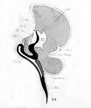
Fig. 24. Diagram of those structures of the central nervous system which lie near the midline and which can be identified. The outline of the subdural space used was obtained from the sagittal section. Posteriorly this passes near to the median margin of the left encephalocele. The cerebrum designated by a dotted line is shown protruding below the foramen magnum into the encephalocele. A small portion of the cerebellum, represented by line-hatching, is seen to lie very much flattened on top of the cord. The brain-stem and cord, much bent, arc shown in solid black. Those cranial nerves which were identified are shown by lines. Only the first spinal nerve is shown. The floor of the fourth ventricle lies inverted on top of a flat cord.
Original plate images magnification shown in brackets.
| Historic Disclaimer - information about historic embryology pages |
|---|
| Pages where the terms "Historic" (textbooks, papers, people, recommendations) appear on this site, and sections within pages where this disclaimer appears, indicate that the content and scientific understanding are specific to the time of publication. This means that while some scientific descriptions are still accurate, the terminology and interpretation of the developmental mechanisms reflect the understanding at the time of original publication and those of the preceding periods, these terms, interpretations and recommendations may not reflect our current scientific understanding. (More? Embryology History | Historic Embryology Papers) |
Glossary Links
- Glossary: A | B | C | D | E | F | G | H | I | J | K | L | M | N | O | P | Q | R | S | T | U | V | W | X | Y | Z | Numbers | Symbols | Term Link
Cite this page: Hill, M.A. (2024, May 2) Embryology Book - Contributions to Embryology Carnegie Institution No.22. Retrieved from https://embryology.med.unsw.edu.au/embryology/index.php/Book_-_Contributions_to_Embryology_Carnegie_Institution_No.22
- © Dr Mark Hill 2024, UNSW Embryology ISBN: 978 0 7334 2609 4 - UNSW CRICOS Provider Code No. 00098G


