Trisomy 21
Introduction
Down syndrome or trisomy 21 is caused by nondisjunction of chromosome 21 in a parent who is chromosomally normal and is one of the most common chromosomal abnormalities in liveborn children. The frequency of trisomy 21 in the population is approximately 1 in 650 to 1,000 live births, in Australia between 1991-97 there were 2,358 Trisomy 21 (Down) infants.
Aneuploidy is the term used to describe any abnormal number of chromosomes either an increase or decrease in total number.
Recent attention has focussed on screening for Down's syndrome (mainly in terms of cost and efficiency) during fetal life with over 350 articles in the medical literature in just the past five years. There is also a high correlation of increased genetic risk with maternal age.
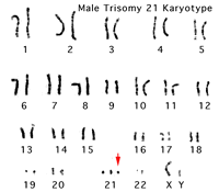
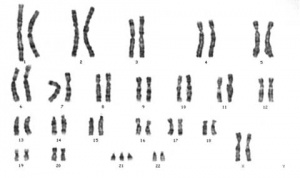
Trisomy 21 (Down Syndrome) Karyotypes
The normal human karyotypes contain 22 pairs of autosomal chromosomes and one pair of sex chromosomes. The karyotype is the characteristic chromosome complement as identified by staining and can only be identified during cell division when chromosomes are folded. The chromosomes when organised as an image in sequence are called a karyogram or idiogram.
Associated Congenital Abnormalities
- neurological (mental retardation)
- characteristic facies
- heart (atrioventricular canal)
- gastrointestinal tract (duodenal stenosis or atresia, imperforate anus, and Hirschsprung disease)
- leukemia (ALL and AML)
- hearing loss (90% of all patients)
- musculoskeletal (limb abnormalities, hypotonia, joint hypermobility, ligamentous laxity, spine anomolies, scoliosis)
- Acute megakaryocytic leukemia occurs 200 to 400 times more frequently in Down syndrome.
Hearing loss is usually of the conductive type. (More? Hearing Abnormalities)
Musculoskeletal include bony anomalies of the cervical spine (produce atlanto-occipital and cervical instability), scoliosis, hip instability, slipped capital femoral epiphysis, patellar instability, and foot deformities. (modified musculoskeletal list from Caird etal., 2006)
Heart Defects
Congenital heart defects are common (40 to 50%) in Down’s babies and are a common cause of postnatal death.
Approximately 30 to 40% have complete atrioventricular septal defects (early diagnosis generally allows corrective surgery to be performed).
- endocardial cushion defect (43%)
- ventricular septal defect (32%)
- secundum atrial septal defect (10%)
- tetralogy of Fallot (6%)
- patent ductus arteriosus (16%)
Limb Defects
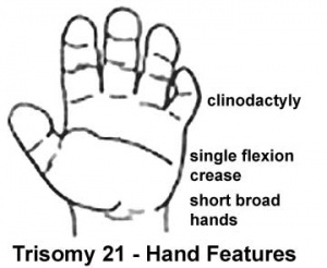
- Hand - features short and broad hands, clinodactyly (curving of the fifth finger, little finger) with a single flexion crease (20%), hyperextensible finger joints.
- Foot - space between the great toe (big) and the second toe is increased.
- Hip - acquired hip dislocation (6%).
Other musculoskeletal effects include bony anomalies of the cervical spine (produce atlanto-occipital and cervical instability), scoliosis, hip instability, slipped capital femoral epiphysis, patellar instability, and foot deformities. PMID: 17030594
(More? Limb Abnormalities)
American College of Obstetricians and Gynecologists Recommendations
The following ACOG recommendations (January 2007) are based on good and consistent scientific evidence:
- First-trimester screening using both nuchal translucency (NT), an ultrasound exam that measures the thickness at the back of the neck of the fetus, and a blood test is an effective screening test in the general population and is more effective than NT alone.
- Women found to be at increased risk of having a baby with Down syndrome with first-trimester screening should be offered genetic counseling and the option of CVS or mid-trimester amniocentesis.
- Specific training, standardization, use of appropriate ultrasound equipment, and ongoing quality assessment are important to achieve optimal NT measurement for Down syndrome risk assessment, and this procedure should be limited to centers and individuals meeting this criteria.
- Neural tube defect screening should be offered in the mid-trimester to women who elect only first-trimester screening for Down syndrome.
Text extract from: New Recommendations for Down Syndrome Call for Screening of All Pregnant Women (press release January 2, 2007)
References
Journals
- Down Syndrome Quarterly DSQ - an interdisciplinary journal devoted to advancing the state of knowledge on Down syndrome
- Down Syndrome Research and Practice Archive Homepage
NCBI Bookshelf
- Modern Genetic Analysis Griffiths, Anthony J.F.; Gelbart, William M.; Miller, Jeffrey H.; Lewontin, Richard C. New York: W. H. Freeman & Co.; c1999. Image - Characteristics of Down syndrome (trisomy 21)
- Introduction to Genetic Analysis 7th ed. Griffiths, Anthony J.F.; Miller, Jeffrey H.; Suzuki, David T.; Lewontin, Richard C.; Gelbart, William M. New York: W. H. Freeman & Co.; c1999. Image - Down syndrome in Robertsonian translocation
- Clinical Methods 3rd ed. Walker, H.K.; Hall, W.D.; Hurst, J.W.; editors Stoneham (MA): Butterworth Publishers; c1990 Table - Recognizable Genetic Conditions
Reviews
- Dent KM, Carey JC. Breaking difficult news in a newborn setting: Down syndrome as a paradigm. Am J Med Genet C Semin Med Genet. 2006 Aug 15;142(3):173-9. PMID: 17048355
- Caird MS, Wills BP, Dormans JP.] Down syndrome in children: the role of the orthopaedic surgeon. J Am Acad Orthop Surg. 2006 Oct;14(11):610-9. PMID: 17048355
- Antonarakis SE, Epstein CJ. The challenge of Down syndrome. Trends Mol Med. 2006 Oct;12(10):473-9. Epub 2006 Aug 28. PMID: 16935027
- Benn PA. Advances in prenatal screening for Down syndrome: II first trimester testing, integrated testing, and future directions. Clin Chim Acta. 2002 Oct;324(1-2):1-11. PMID: 12204419
- Maymon R, Jauniaux E. Down's syndrome screening in pregnancies after assisted reproductive techniques: an update. Reprod Biomed Online. 2002 May-Jun;4(3):285-93. PMID: 12709282
- Souter VL, Nyberg DA. Sonographic screening for fetal aneuploidy: first trimester. J Ultrasound Med. 2001 Jul;20(7):775-90. PMID: 11444737
- Jackson M, Rose NC. Diagnosis and management of fetal nuchal translucency. Semin Roentgenol. 1998 Oct;33(4):333-8. Review. PMID: 9800243
- Menendez M. Down syndrome, Alzheimer's disease and seizures. Brain Dev. 2005 Jun;27(4):246-52. PMID: 15862185
- FitzPatrick DR. Transcriptional consequences of autosomal trisomy: primary gene dosage with complex downstream effects. Trends Genet. 2005 May;21(5):249-53. PMID: 15851056
Articles
- Van Riper M.] Van Riper M. Families of children with down syndrome: responding to "a change in plans" with resilience. J Pediatr Nurs. 2007 Apr;22(2):116-28. PMID: 17382849
- Malone FD, Canick JA, Ball RH, Nyberg DA, Comstock CH, Bukowski R, Berkowitz RL, Gross SJ, Dugoff L, Craigo SD, Timor-Tritsch IE, Carr SR, Wolfe HM, Dukes K, Bianchi DW, Rudnicka AR, Hackshaw AK, Lambert-Messerlian G, Wald NJ, D'Alton ME. First-Trimester or Second-Trimester Screening, or Both, for Down's Syndrome. N Engl J Med. 2005 Nov 10;353(19):2001-2011. PMID: 16282175 A large team of clinical researchers have compared the effectiveness of first and second trimester screening methods for this chromosome 21 trisomy disorder
- "First-trimester combined screening at 11 weeks of gestation is better than second-trimester quadruple screening but at 13 weeks has results similar to second-trimester quadruple screening. Both stepwise sequential screening and fully integrated screening have high rates of detection of Down's syndrome, with low false positive rates."
First-trimester combined screening - nuchal translucency, pregnancy-associated plasma protein A [PAPP-A], free beta subunit of hCG (10 weeks 3 days through 13 weeks 6 days of gestation) Second-trimester quadruple screening - alpha-fetoprotein, total hCG, unconjugated estriol, and inhibin A at (15 through 18 weeks of gestation). (NEMJ Nov 10) NEMJ - Down's Syndrome Screening Article
Associated Neurological
- Menendez M. Down syndrome, Alzheimer's disease and seizures. Brain Dev. 2005 Jun;27(4):246-52. PMID: 15862185
- "Neuropathologically, Alzheimer-type abnormalities are demonstrated in patients with Down syndrome (DS), both demented and nondemented and more than a half of patients with DS above 50 years develop Alzheimer's disease (AD)."
- Malone FD, Canick JA, Ball RH, Nyberg DA, Comstock CH, Bukowski R, Berkowitz RL, Gross SJ, Dugoff L, Craigo SD, Timor-Tritsch IE, Carr SR, Wolfe HM, Dukes K, Bianchi DW, Rudnicka AR, Hackshaw AK, Lambert-Messerlian G, Wald NJ, D'Alton ME. First-Trimester or Second-Trimester Screening, or Both, for Down's Syndrome. N Engl J Med. 2005 Nov 10;353(19):2001-2011. PMID: 16282175
Hook EB, Cross PK, Schreinemachers DM.Chromosomal abnormality rates at amniocentesis and in live-born infants. JAMA. 1983 Apr 15;249(15):2034-8.
Schreinemachers DM, Cross PK, Hook EB. Rates of trisomies 21, 18, 13 and other chromosome abnormalities in about 20 000 prenatal studies compared with estimated rates in live births. Hum Genet. 1982;61(4):318-24.
New triple screen test for Down syndrome: combined urine analytes and serum AFP. Bahado-Singh RO, et al.J Matern Fetal Med. 1998 May-Jun;7(3):111-4.
Screening for Down's syndrome: effects, safety, and cost effectiveness of first and second trimester strategies R E Gilbert, C Augood, R Gupta, A E Ades, S Logan, M Sculpher, J H P van der Meulen, Euan M Wallace, and Sheila Mulvey BMJ 2001; 323: 423 (link to paper)
Noninvasive means of identifying fetuses with possible Down syndrome: a review. Kubas C. J Perinat Neonatal Nurs 1999 Sep;13(2):27-46 Women who are 35 years or older are offered invasive prenatal testing because of the increased risk of chromosomal abnormalities, especially Down syndrome. In an attempt to increase the number of Down syndrome fetuses being detected and decrease the number of invasive procedures being performed on pregnancies not affected with a chromosome abnormality, both biochemical and ultrasound screening methods are being studied and are summarized in this article.
The ultrasound markers reviewed include increased nuchal thickness, increased nuchal lucency, shortened femur, shortened humerus, pyelectasis, hypoplastic ears, echogenic intracardiac focus, hypoplasia of the fifth middle phalanx, and echogenic bowel.
Books
Note: books are listed for educational and information purposes only and does not suggest a commercial product endorsement.
Search PubMed
Search PubMed Now: Trisomy 21 | Down Syndrome | aneuploidy |
WWW Links
OMIM Down Syndrome
Trisomy Organization http://www.trisomy.org/
Better Health Victoria trisomy disorders
See also Virtual Hospital male Karyotype (Virtual Hospital now inactive).
The Australian Down Syndrome Association Inc
c/o - Down Syndrome Association of NSW IncPO Box 2356 (31 O'Connell Street)North Parramtta NSW 2151 AustraliaTel. 02 9683 4333 Fax. 02 9683 420E-mail:dsansw@hartingdale.com.au
The ACT Down Syndrome Association
P.O. Box 717Mawson ACT 2607Tel: 06 290 1984 Fax: 06 286 4475Email: ehoek@pcug.org.au
The Down Syndrome Association of QueenslandP.O. Box 1293Milton Queensland 4064
UNSW Embryology Links
Terms
alpha-fetoprotein - (AFP) A serum fetal glycoprotein produced by both the yolk sac and fetal liver. The presence of the protein in maternal blood is the basis of a test for genetic or developmental problems in the fetus. Low levels of AFP normally occur in the blood of a pregnant woman, high levels may indicate neural tube defects (spina bifida, anencephaly). (More? Abnormal Development- AFP test)
alpha-fetoprotein test (APF test) A prenatal test to measure the amount of a fetal protein in the mother's blood (or amniotic fluid). Abnormal amounts of the protein may indicate genetic or developmental problems in the fetus. Serum alpha-fetoprotein (AFP) is a fetal glycoprotein produced by the yolk sac and fetal liver. Low levels of AFP normally occur in the blood of a pregnant woman, high levels may indicate neural tube defects (spina bifida, anencephaly). (More? Abnormal Development- AFP test)
aneuploidy - Term used to describe an abnormal number of chromosomes mainly (90%) due to chromosome malsegregation mechanisms in maternal meiosis I. (More? Meiosis)
karyotype - (Greek, karyon = kernel or nucleus + typos = stamp) Term used to describe the chromosomal (genetic) makeup (complement) of a cell. (More? Week 1 Notes | Genetic Abnormalities)
single umbilical artery - (SUA) Placental cord with only a single placental artery (normally paired). This abnormality can be detected by ultrasound (colour flow imaging of the fetal pelvis) and is used as an indicator for further prenatal diagnostic testing for chromosomal abnormalities and other systemic defects. (More? Prenatal Diagnosis | Ultrasound)
trimester - Clinical term used to describe and divide human pregnancy period (9 months) into three equal parts of about three calendar months. The first trimester corresponds approximately to embryonic development (week 1 to 8) of organogenesis and early fetal. The second and third trimester correspond to the fetal period of growth in size (second trimester) and weight (third trimester), as well as continued differentiation of existing organs and tissues. (More? Embryo Stages | Human Fetal Period | Development Week by Week)
Glossary Links
A | B | C | D | E | F | G | H | I | J | K | L | M | N | O | P | Q | R | S | T | U | V | W | X | Y | Z
- Dr Mark Hill 2009, UNSW Embryology ISBN: 978 0 7334 2609 4 - UNSW CRICOS Provider Code No. 00098G


