Talk:Museum of Natural History Berlin - 2013 Seminar: Difference between revisions
From Embryology
mNo edit summary |
mNo edit summary |
||
| Line 11: | Line 11: | ||
===Animal Models=== | ===Animal Models=== | ||
* Experimental embryology - involving manipulation of animal models. | |||
* Imaging development - electron microscopy, fluorescence microscopy, confocal microscopy, ultrasound, tomography. | |||
* Research fields - molecular biology, in vitro fertilisation, stem cells. | |||
{| | {| | ||
| [[File:Chick E12.jpg|100px]] | | [[File:Chick E12.jpg|100px]] | ||
| Line 27: | Line 31: | ||
|} | |} | ||
==Education | ==Recent Embryology Education== | ||
===Teaching Contact Hours=== | |||
{| | |||
| [[File:Anatomy teaching USA graph.jpg|600px]] | |||
Trends in anatomy disciplines contact teaching hours based on data from USA survey.<ref><pubmed>19890982</pubmed></ref> | |||
| {{Human development movie 1}} | |||
|} | |||
{| class="wikitable collapsible collapsed" | |||
! About this graph | |||
|- | |||
| | |||
* Online survey were constructed to gather basic information about gross anatomy, microscopic anatomy, neuroscience/neuroanatomy, and embryology courses. | |||
* 2009 total of 130 allopathic and 25 osteopathic medical schools in USA. | |||
* Number of responses (Gross anatomy 65, Microscopic anatomy 45, Neuroscience/Neuroanatomy 31, Embryology 43) | |||
'''Reference''' | |||
<references /> | |||
|} | |||
===Embryo Serial Sections=== | ===Embryo Serial Sections=== | ||
{| | {| | ||
| Line 36: | Line 59: | ||
View embryo section 49 images (7x7 matrix) in 3 sets: | View embryo section 49 images (7x7 matrix) in 3 sets: | ||
# | # [[Carnegie_stage_13_-_serial_sectionsStage 13 Embryo]] (mid-embryonic period) | ||
# Stage 22 Embryo | # [[Carnegie_stage_22_-_serial_sections|Stage 22 Embryo]] (end embryonic period) | ||
# Selected Stage 22 images ( | # [[Carnegie stage 22 - selected serial sections|Selected Stage 22 images]] (selected organs and tissues from end embryonic) | ||
Separate sets of labelled drawing corresponding to each section. | Separate sets of labelled drawing corresponding to each section. | ||
|} | |} | ||
{| class="wikitable collapsible collapsed" | {| class="wikitable collapsible collapsed" | ||
! | ! Microfiche Images | ||
|- | |- | ||
| {{Stage13ULicon120}} | | {{Stage13ULicon120}} | ||
Revision as of 00:40, 25 November 2013
Good morning, I apologise that my talk today will not be in German.
Gutenmorgen, entschuldige mich ich, dass mein Gespräch heute nicht auf Deutsch ist.
| Collapse template |
|---|
Animal Models
- Experimental embryology - involving manipulation of animal models.
- Imaging development - electron microscopy, fluorescence microscopy, confocal microscopy, ultrasound, tomography.
- Research fields - molecular biology, in vitro fertilisation, stem cells.

|

|

|

|

|

|
| Chicken Development | Frog Development | Rat Development | Mouse Development | Fly Development | Zebrafish Development |
Recent Embryology Education
Teaching Contact Hours

Trends in anatomy disciplines contact teaching hours based on data from USA survey.[1] |
|
| About this graph |
|---|
Reference
|
Embryo Serial Sections

|
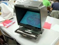
Microfiche Reader used in Medicine and Science classes from 1975 to 1995.
View embryo section 49 images (7x7 matrix) in 3 sets:
Separate sets of labelled drawing corresponding to each section. |
| Microfiche Images | ||||||||||||||||||||||||||||||||||||||||||||||||||||||||||||||||||||||||||||||||||||||||||||||||||
|---|---|---|---|---|---|---|---|---|---|---|---|---|---|---|---|---|---|---|---|---|---|---|---|---|---|---|---|---|---|---|---|---|---|---|---|---|---|---|---|---|---|---|---|---|---|---|---|---|---|---|---|---|---|---|---|---|---|---|---|---|---|---|---|---|---|---|---|---|---|---|---|---|---|---|---|---|---|---|---|---|---|---|---|---|---|---|---|---|---|---|---|---|---|---|---|---|---|---|
| ||||||||||||||||||||||||||||||||||||||||||||||||||||||||||||||||||||||||||||||||||||||||||||||||||
| ||||||||||||||||||||||||||||||||||||||||||||||||||||||||||||||||||||||||||||||||||||||||||||||||||
|


1997 Online
pre-1997

2001
2004
2007
2007 DVD


| Technology Timeline | |
|---|---|

The changing technology environment we have experienced in the last 50 years, and more recently in the last 20 "internet years". | |
|
|