File list
From Embryology
This special page shows all uploaded files.
| Date | Name | Thumbnail | Size | User | Description | Versions |
|---|---|---|---|---|---|---|
| 01:40, 17 November 2013 | Ziegler model 32.jpg (file) |  |
41 KB | Z8600021 | ==Ziegler Model - Development History of the Eye - Series 8c Model 1== {{Ziegler Eye Models}} | 1 |
| 01:51, 17 November 2013 | Ziegler model 31.jpg (file) |  |
0 bytes | Z8600021 | ==Ziegler Model - Development History of the Eye - Series 8c Model 1== {{Ziegler Eye Models}} | 1 |
| 01:38, 17 November 2013 | Ziegler model 30.jpg (file) |  |
30 KB | Z8600021 | ==Ziegler Model - Development History of the Eye - Series 8c Model 1== {{Ziegler Eye Models}} | 1 |
| 00:42, 17 November 2013 | Ziegler model 29.jpg (file) |  |
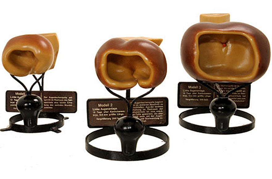
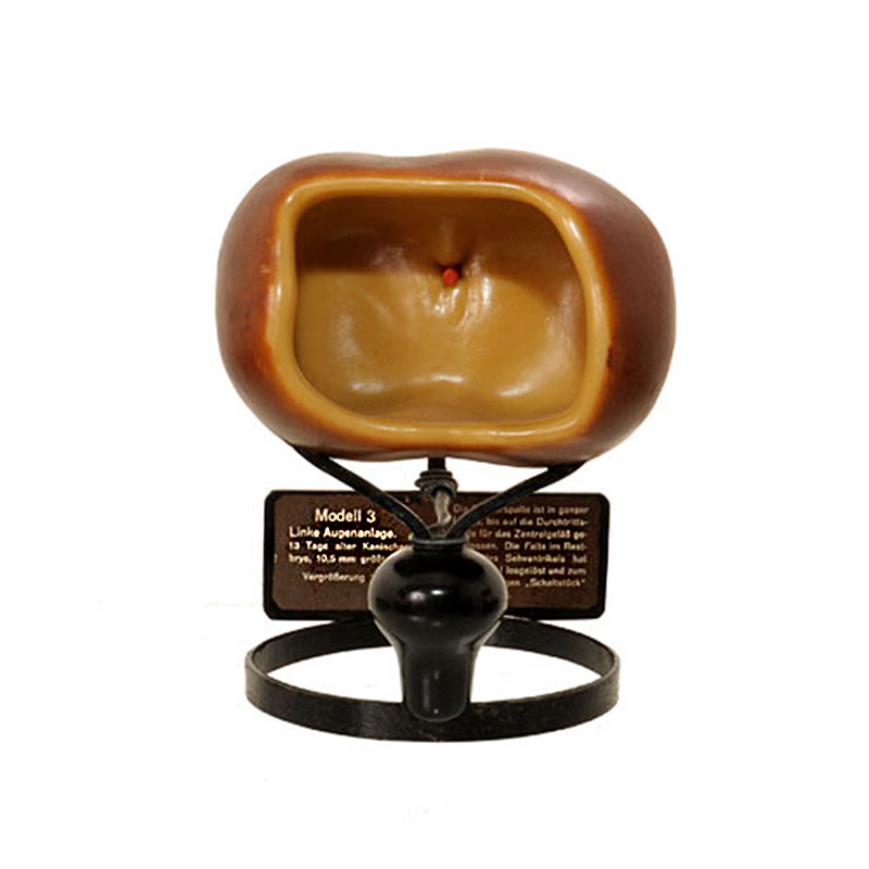
69 KB | Z8600021 | ==Ziegler Model - Development History of the Eye - Series 8b== Model 3 - Primordial eye of 13 days old embryo 10.5 mm greatest length (CRL), external view. {{Ziegler Eye Models}} | 1 |
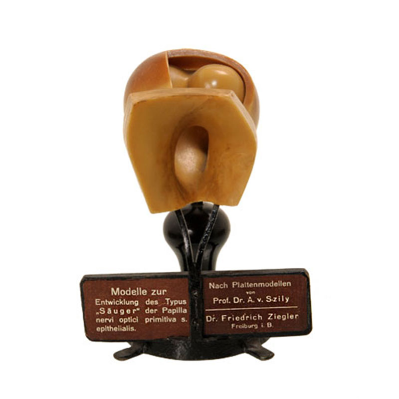
| 00:34, 17 November 2013 | Ziegler model 28.jpg (file) |  |
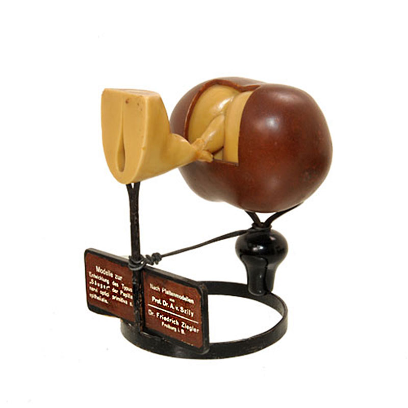
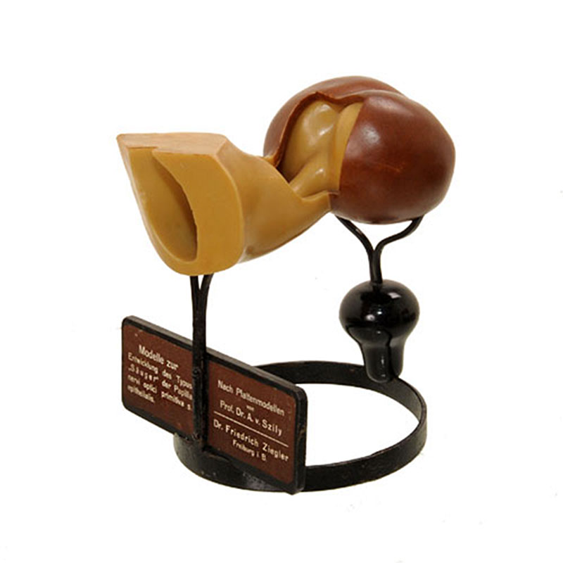
49 KB | Z8600021 | ==Ziegler Model - Development History of the Eye - Model 3== Primordial eye of 13 days old embryo 10.5 mm greatest length (CRL), lateral view. See also model legend detail image. {{Ziegler Eye Models}} | 1 |
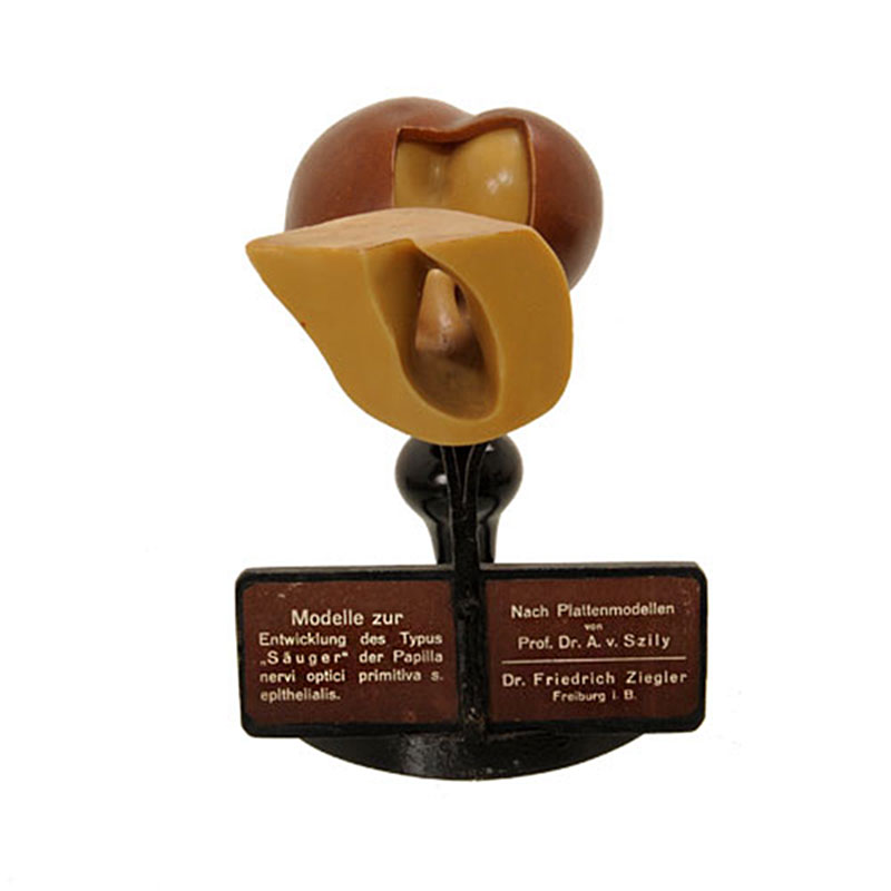
| 00:31, 17 November 2013 | Ziegler model 27.jpg (file) |  |
52 KB | Z8600021 | ==Ziegler Model - Development History of the Eye - Model 3== Primordial eye of 13 days old embryo 10.5 mm greatest length (CRL), external view. See also model legend detail image. {{Ziegler Eye Models}} | 1 |
| 00:21, 17 November 2013 | Ziegler model 26.jpg (file) |  |
48 KB | Z8600021 | ==Ziegler Model - Development History of the Eye - Model 3== Primordial eye of 13 days old embryo 9.6 mm greatest length (CRL), external view. See also model legend detail image. {{Ziegler Eye Models}} | 1 |
| 00:14, 17 November 2013 | Ziegler model 25.jpg (file) |  |
46 KB | Z8600021 | ==Ziegler Model - Development History of the Eye - Model 2== Primordial eye of 13 days old embryo 9.6 mm greatest length (CRL), lateral view. See also model legend detail image. {{Ziegler Eye Models}} | 1 |
| 00:12, 17 November 2013 | Ziegler model 24.jpg (file) |  |
43 KB | Z8600021 | ==Ziegler Model - Development History of the Eye - Model 2== Primordial eye of 13 days old embryo 9.6 mm greatest length (CRL), internal view. See also model legend detail image. {{Ziegler Eye Models}} | 1 |
| 00:01, 17 November 2013 | Ziegler model 23.jpg (file) |  |
49 KB | Z8600021 | ==Ziegler Model - Development History of the Eye - Model 2== Left primordial eye of 12 days old embryo, external view. See also model legend detail image. {{Ziegler Eye Models}} | 1 |
| 23:48, 16 November 2013 | Ziegler model 22.jpg (file) |  |
41 KB | Z8600021 | ==Ziegler Model - Development History of the Eye - Model 1== Left primordial eye of 12 days old embryo, lateral view. See also model legend detail image. {{Ziegler Eye Models}} | 1 |
| 23:18, 16 November 2013 | Ziegler model 21.jpg (file) |  |
44 KB | Z8600021 | ==Ziegler Model - Development History of the Eye - Model 1== See also model legend detail image. {{Ziegler Eye Models}} | 1 |
| 22:25, 16 November 2013 | Ziegler model 20.jpg (file) |  |
38 KB | Z8600021 | ==Ziegler Model== {{Ziegler Models}} | 1 |
| 12:41, 8 May 2012 | Ziegler model 19.jpg (file) |  |
23 KB | Z8600021 | ==Ziegler Heart Model== {{Ziegler Models}} Category:Heart | 1 |
| 12:38, 8 May 2012 | Ziegler model 18.jpg (file) |  |
42 KB | Z8600021 | ==Ziegler Heart Model== {{Ziegler Models}} Category:Heart | 1 |
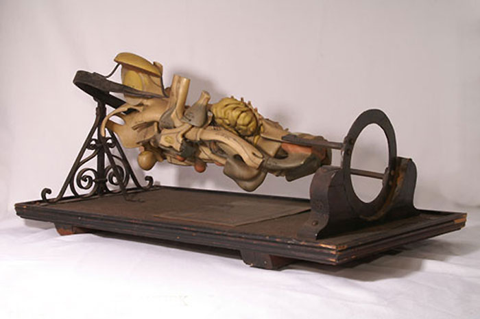
| 12:32, 8 May 2012 | Ziegler model 17.jpg (file) |  |
41 KB | Z8600021 | ==Ziegler Medulla Model== {{Ziegler Models}} | medulla model 1 | medulla model 2 | medulla model 3 | medulla model 4 |[[Neural_-_Medulla_Obl | 1 |
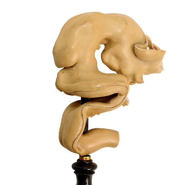
| 12:26, 8 May 2012 | Ziegler model 16.jpg (file) |  |
56 KB | Z8600021 | ==Ziegler Medulla Model== {{Ziegler Models}} | medulla model 1 | medulla model 2 | Medulla Category:Neural | 1 |
| 12:01, 8 May 2012 | Ziegler model 15.jpg (file) |  |
61 KB | Z8600021 | ==Ziegler Model Information== Image of information attached to underside of model base. {{Ziegler Models}} | 1 |
| 11:57, 8 May 2012 | Ziegler model 14.jpg (file) |  |
30 KB | Z8600021 | 2 | |
| 11:54, 8 May 2012 | Ziegler model 13.jpg (file) |  |
30 KB | Z8600021 | ==Ziegler Brain Model== {{Ziegler Models}} Category:Neural | 1 |
| 11:51, 8 May 2012 | Ziegler model 12.jpg (file) |  |
39 KB | Z8600021 | ==Ziegler Heart Model== {{Ziegler Models}} Category:Heart | 1 |
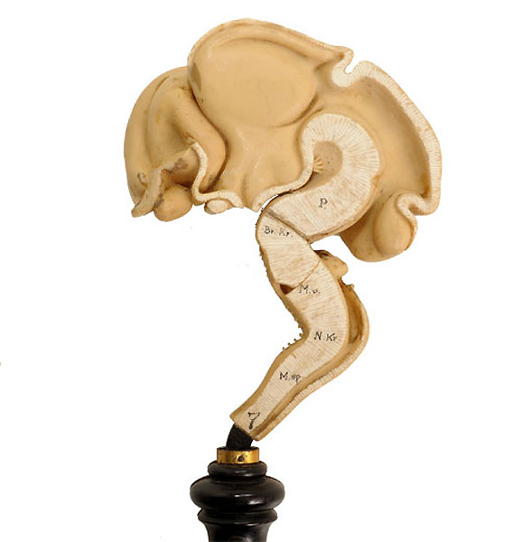
| 11:47, 8 May 2012 | Ziegler model 11.jpg (file) |  |
43 KB | Z8600021 | ==Ziegler Medulla Model== {{Ziegler Models}} | Medulla Category:Neural | 1 |
| 11:43, 8 May 2012 | Ziegler model 10.jpg (file) |  |
41 KB | Z8600021 | ==Ziegler Medulla Model== {{Ziegler Models}}| Medulla Category:Neural | 1 |
| 11:38, 8 May 2012 | Ziegler model 09.jpg (file) |  |
30 KB | Z8600021 | ==Ziegler Brain Model== {{Ziegler Models}} Category:Neural | 1 |
| 11:36, 8 May 2012 | Ziegler model 08.jpg (file) |  |
33 KB | Z8600021 | ==Ziegler Brain Model== {{Ziegler Models}} | 1 |
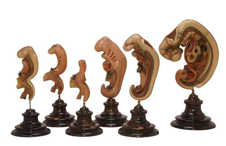
| 11:21, 8 May 2012 | Ziegler model 07.jpg (file) |  |
58 KB | Z8600021 | ==Ziegler Models== {{Ziegler Models}} | 1 |
| 11:18, 8 May 2012 | Ziegler model 06.jpg (file) |  |
37 KB | Z8600021 | ==Ziegler Model== {{Ziegler Models}} | 1 |
| 12:51, 8 May 2012 | Ziegler model 05.jpg (file) |  |
43 KB | Z8600021 | 1 | |
| 11:09, 8 May 2012 | Ziegler model 04.jpg (file) |  |
36 KB | Z8600021 | ==Ziegler Models== {{Ziegler_Models}} | 1 |
| 11:05, 8 May 2012 | Ziegler model 03.jpg (file) |  |
31 KB | Z8600021 | ==Ziegler Model== {{Ziegler Models}} | 1 |
| 10:46, 8 May 2012 | Ziegler model 02.jpg (file) |  |
29 KB | Z8600021 | {{Ziegler_Models}} | 1 |
| 10:37, 8 May 2012 | Ziegler model 01.jpg (file) |  |
43 KB | Z8600021 | ==Ziegler Model== {{Ziegler Models}} | 1 |
| 23:28, 23 November 2013 | Ziegler Freiburg workshop.jpg (file) |  |
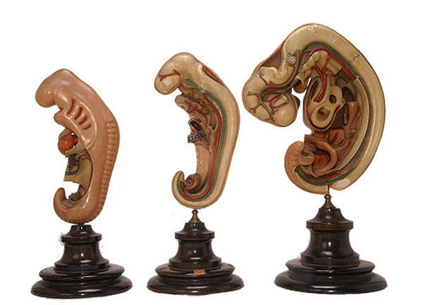
104 KB | Z8600021 | ==Ziegler Freiburg workshop== Photograph of Ziegler (left foreground) in the Ziegler Freiburg workshop (c 1912). :Links: Ziegler Models Source: Photograph from Friedrich Ziegler, Embryologische Wachsmodelle,... | 1 |
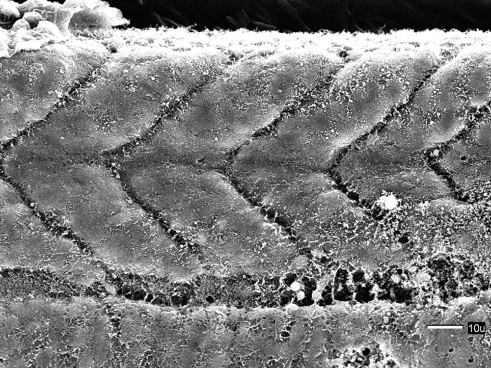
| 14:10, 12 December 2014 | Zebrafish trunk SEM02.jpg (file) |  |
163 KB | Z8600021 | 1 | |
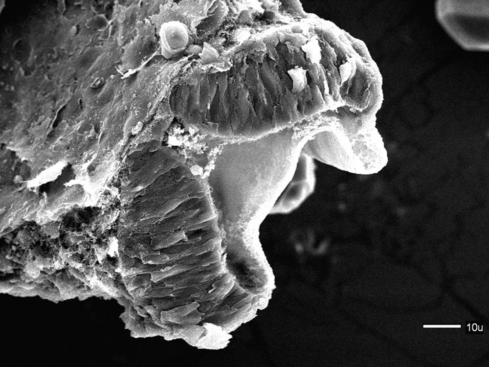
| 14:04, 12 December 2014 | Zebrafish trunk SEM01.jpg (file) |  |
222 KB | Z8600021 | ==Zebrafish trunk (day 1) SEM== Scanning EM of a 24 hr (prim-5) zebrafish embryo. {{Zebrafish SEM links}} ===Reference=== Zebrafish were chemically fixed critically point dried, and sputter coated with gold/palladium. This image is part of a serie... | 1 |
| 09:47, 16 November 2012 | Zebrafish skull neural crest.jpg (file) |  |
187 KB | Z8600021 | ==Zebrafish Skull Neural Crest== ===Reference=== Kague E, Gallagher M, Burke S, Parsons M, Franz-Odendaal T, et al. (2012) Skeletogenic Fate of Zebrafish Cranial and Trunk Neural Crest. PLoS ONE 7(11): e47394. doi:10.1371/journal.pone.0047394 [http://w | 1 |
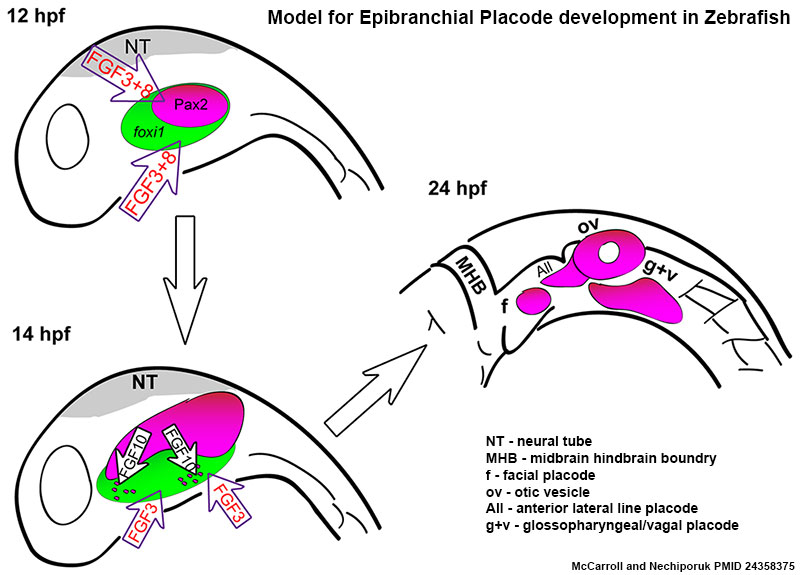
| 00:32, 27 December 2013 | Zebrafish placode model.jpg (file) |  |
82 KB | Z8600021 | ==Zebrafish Epibrachial Placode Model== Before 12 hpf, Fgf3 and Fgf8 specify cells of the posterior placodal area that will give rise to the otic and EB placodes. Shortly after, between 14 and 22 hpf, Fgf10a, expressed by the anterior lateral line pre... | 1 |
| 14:17, 12 December 2014 | Zebrafish perichordal sheath SEM.jpg (file) |  |
117 KB | Z8600021 | ==Zebrafish perichordal sheath (day 1) SEM== Scanning EM of a 24 hr (prim-5) zebrafish embryo. {{Zebrafish SEM links}} ===Reference=== <pubmed>15602926</pubmed> Specimens were chemically fixed critically point dried, and sputter coated with gold/p... | 1 |
| 16:02, 14 October 2012 | Zebrafish neural crest model.jpg (file) |  |
61 KB | Z8600021 | ==Model for the role of H3.3-dependent histone replacement during CNC development== At the early embryonic blastula stage, cells have a broad potential with cis-regulatory elements for developmental genes existing in a “poised” chromatin state. After | 1 |
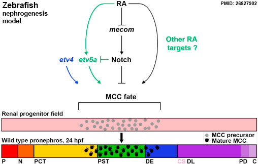
| 10:22, 31 August 2016 | Zebrafish nephrogenesis signaling01.jpg (file) |  |
29 KB | Z8600021 | ==Zebrafish nephrogenesis signaling== ===Reference=== <pubmed>26827902</pubmed> ====Copyright==== https://creativecommons.org/licenses/by/4.0/ Fig. 7. 1-s2.0-S0012160615301068-gr7.jpg PMID and title added to original | 1 |
| 14:02, 12 December 2014 | Zebrafish myotomes SEM.jpg (file) |  |
250 KB | Z8600021 | ==Zebrafish myotomes (day 1) SEM== Scanning EM of a 24 hr (prim-5) zebrafish embryo. {{Zebrafish SEM links}} ===Reference=== Zebrafish were chemically fixed critically point dried, and sputter coated with gold/palladium. This image is part of a se... | 1 |
| 15:08, 7 August 2015 | Zebrafish movie01.mp4 (file) | 10.48 MB | Z8600021 | 1 | ||
| 14:46, 20 February 2011 | Zebrafish melanocyte development model.jpg (file) |  |
40 KB | S8600021 | ==Model for the parallel establishment of the zebrafish embryonic melanocyte lineage and the adult melanocyte stem cell lineage== Our results indicate that there are two distinct melanocyte lineages that develop in the zebrafish embryo: the embryonic or | 1 |
| 14:36, 3 September 2009 | Zebrafish image.jpg (file) |  |
39 KB | Z3223194 | Original image source: http://en.wikipedia.org/wiki/File:Zebrafisch.jpg The copyright holder of this work allows anyone to use it for any purpose including unrestricted redistribution, commercial use, and modification. | 1 |
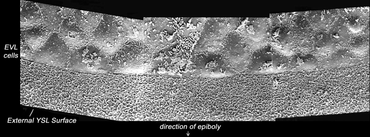
| 14:30, 12 December 2014 | Zebrafish enveloping layer SEM02.jpg (file) |  |
117 KB | Z8600021 | 1 | |
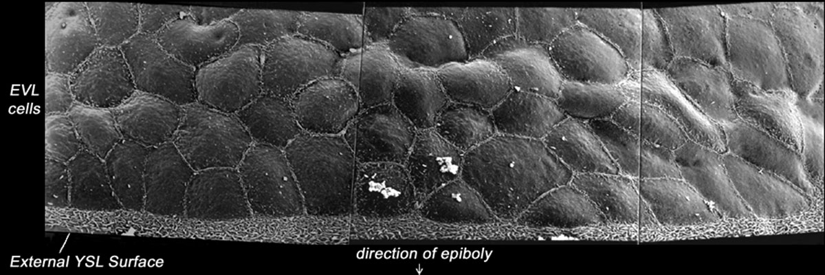
| 14:29, 12 December 2014 | Zebrafish enveloping layer SEM01.jpg (file) |  |
150 KB | Z8600021 | 1 | |
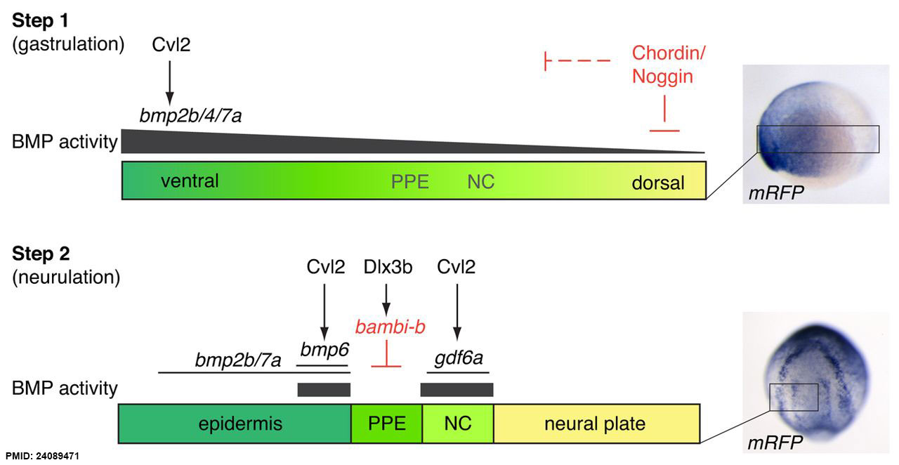
| 22:44, 3 March 2015 | Zebrafish ectodermal patterning model.jpg (file) |  |
80 KB | Z8600021 | A two-step model for zebrafish ectodermal patterning. Step 1 depicts the DV gradient present during gastrulation. High BMP activity is observed ventrally as generated by bmp2b/4/7a ligand expression and reinforced by Cvl2. BMP activity is inhibited dor... | 1 |
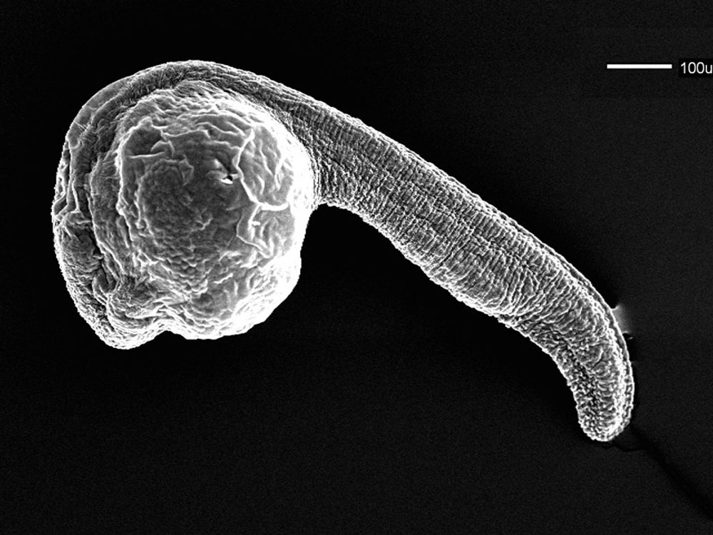
| 08:16, 26 April 2011 | Zebrafish day 1 SEM.jpg (file) |  |
130 KB | S8600021 | ==Zebrafish Day 1 SEM== Scanning EM of a 24 hr (prim-5) zebrafish embryo. Zebrafish were chemically fixed critically point dried, and sputter coated with gold/palladium. This image is part of a series taken by Bryan Crawford while he was at the Univer | 1 |
| 08:23, 26 April 2011 | Zebrafish brain fold SEM.jpg (file) |  |
172 KB | S8600021 | ==Zebrafish Brain Fold SEM== Scanning EM of the zebrafish head folds. Specimens were chemically fixed critically point dried, and sputter coated with gold/palladium. This image is part of a series taken by [http://www.unb.ca/fredericton/science/biology | 1 |
| 09:05, 4 December 2014 | Zebrafish Wdr18 expression 02.jpg (file) |  |
69 KB | Z8600021 | 1 |