Smooth Muscle Development: Difference between revisions
mNo edit summary |
mNo edit summary |
||
| Line 13: | Line 13: | ||
|-bgcolor="F5FAFF" | |-bgcolor="F5FAFF" | ||
| | | | ||
* '''Smooth muscle cell differentiation in vitro: models and underlying molecular mechanisms'''<ref><pubmed>21677291</pubmed></ref> "Development of in vitro models by which to study smooth muscle cell (SMC) differentiation has been hindered by some peculiarities intrinsic to these cells, namely their different embryological origins and their ability to undergo phenotypic modulation in cell culture. Although many in vitro models are available for studying SMC differentiation, careful consideration should be taken so that the model chosen fits the questions being posed. In this review, we summarize several well-established in vitro models available to study SMC differentiation from stem cells and outline novel mechanisms recently identified as underlying SMC differentiation programs." | * '''Prdm6 is essential for cardiovascular development in vivo'''<ref name=PMID24278461><pubmed>24278461</pubmed></ref> "Complete deletion of Prdm6 results in embryonic lethality due to cardiovascular defects associated with aberrations in vascular patterning. However, smooth muscle cells could be regularly differentiated from Prdm6-deficient embryonic stem cells and vascular smooth muscle cells were present and proliferated normally in Prdm6-deficient embryos. Conditional deletion of Prdm6 in the smooth muscle cell lineage using a SM22-Cre driver line resulted in perinatal lethality due to hemorrhage in the lungs. We thus identified Prdm6 as a factor that is essential for the physiological control of cardiovascular development." | ||
* '''Smooth muscle cell differentiation in vitro: models and underlying molecular mechanisms'''<ref name=PMID21677291><pubmed>21677291</pubmed></ref> "Development of in vitro models by which to study smooth muscle cell (SMC) differentiation has been hindered by some peculiarities intrinsic to these cells, namely their different embryological origins and their ability to undergo phenotypic modulation in cell culture. Although many in vitro models are available for studying SMC differentiation, careful consideration should be taken so that the model chosen fits the questions being posed. In this review, we summarize several well-established in vitro models available to study SMC differentiation from stem cells and outline novel mechanisms recently identified as underlying SMC differentiation programs." | |||
|} | |} | ||
{| class="wikitable collapsible collapsed" | {| class="wikitable collapsible collapsed" | ||
Revision as of 00:04, 2 December 2013
Introduction

There are 3 different types of muscle: skeletal, cardiac and smooth. This page describes smooth muscle development, descriptions of cardiac muscle and smooth muscle development can be found in other notes.
- Smooth muscle is mesoderm in origin and contributes to many different tissues including the muscular wall of the gastrointestinal tract, respiratory tract, artery walls, bladder wall, uterus, seminiferous tubules and ductus deferens.
- Smooth muscle is non-striated in appearance, lacking the regular organisation of sarcomeres seen in skeletal and cardiac muscle.
- Smooth Muscle Links: Smooth Muscle Development | Smooth Muscle Histology | Blood Vessel | Uterus | Urinary Bladder | Mesoderm
Some Recent Findings
|
| More recent papers |
|---|
 This table allows an automated computer search of the external PubMed database using the listed "Search term" text link.
More? References | Discussion Page | Journal Searches | 2019 References | 2020 References Search term: Smooth Muscle Embryology <pubmed limit=5>Smooth Muscle Embryology</pubmed> |
Gastrointestinal Tract
The gastrointestinal tract consists of two thick outer muscle layers (longitudinal and circular) and a thin muscularis mucosa layer.
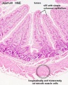
Jejunum
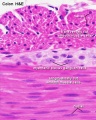
Colon

Neural Innervation
- 1857 Meissner was the first to describe a nerve plexus in the submucosa of the bowel wall.
- 1864 Auerbach described the myenteric plexus between the longitudinal and circular muscle layers.
- 1981 LeDouarin describes neural crest contribution to both plexuses.
Myenteric Plexus
- Auerbach's Plexus
- functions for peristalsis and gastric mixing.
- Coordinated waves of descending inhibition followed by waves of descending excitation
- + Extrinsic parasympathetic cholinergic nerves (vagal and sacral) excite peristalsis and stimulate
- - Sympathetic noradrenergic nerves inhibit the transit of gut contents
Submucosal Plexus
- Meissner's Plexus
- functions for secretion and absorption.
Interstitial cells of Cajal
- Interstitial cells of Cajal (ICCs) are peripheral nervous system neuron found in some smooth muscle organs.
- function as pacemaker cells, neuromodulation or mechanosensory roles.
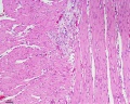
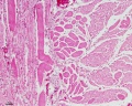
Histology Images
- Smooth Muscle Histology: Labeled Colon low | Labeled Colon high | Colon x40 | Colon x40 | Ileum x10 | Oesophagus x10 | Seminiferous tubule x40 | Uterus myometrium x10 | Uterus myometrium x40 |
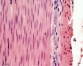
Colon x40
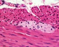
Colon x40
Ileum x10
Oesophagus x10
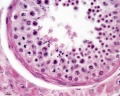
Seminiferous tubule x40
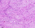
Uterus myometrium x10
Uterus myometrium x40

Ductus deferens


References
Search PubMed
Search Pubmed Central Images: smooth muscle
Search Pubmed: Smooth Muscle Development
Additional Images
Terms
External Links
External Links Notice - The dynamic nature of the internet may mean that some of these listed links may no longer function. If the link no longer works search the web with the link text or name. Links to any external commercial sites are provided for information purposes only and should never be considered an endorsement. UNSW Embryology is provided as an educational resource with no clinical information or commercial affiliation.
Glossary Links
- Glossary: A | B | C | D | E | F | G | H | I | J | K | L | M | N | O | P | Q | R | S | T | U | V | W | X | Y | Z | Numbers | Symbols | Term Link
Cite this page: Hill, M.A. (2024, May 1) Embryology Smooth Muscle Development. Retrieved from https://embryology.med.unsw.edu.au/embryology/index.php/Smooth_Muscle_Development
- © Dr Mark Hill 2024, UNSW Embryology ISBN: 978 0 7334 2609 4 - UNSW CRICOS Provider Code No. 00098G