Paper - Some Observations on the Development of the Ventral Pancreas in Man
| Embryology - 26 Apr 2024 |
|---|
| Google Translate - select your language from the list shown below (this will open a new external page) |
|
العربية | català | 中文 | 中國傳統的 | français | Deutsche | עִברִית | हिंदी | bahasa Indonesia | italiano | 日本語 | 한국어 | မြန်မာ | Pilipino | Polskie | português | ਪੰਜਾਬੀ ਦੇ | Română | русский | Español | Swahili | Svensk | ไทย | Türkçe | اردو | ייִדיש | Tiếng Việt These external translations are automated and may not be accurate. (More? About Translations) |
Odgers PN. Some observations on the development of the ventral pancreas in man. (1930) J. Anat., 65(1): 1-7. PMID 17104298
| Historic Disclaimer - information about historic embryology pages |
|---|
| Pages where the terms "Historic" (textbooks, papers, people, recommendations) appear on this site, and sections within pages where this disclaimer appears, indicate that the content and scientific understanding are specific to the time of publication. This means that while some scientific descriptions are still accurate, the terminology and interpretation of the developmental mechanisms reflect the understanding at the time of original publication and those of the preceding periods, these terms, interpretations and recommendations may not reflect our current scientific understanding. (More? Embryology History | Historic Embryology Papers) |
Some Observations on the Development of the Ventral Pancreas in Man
By P. N. B. Odgers
Department of Human Anatomy, Oxford University
Since 1888, when Phisalix first demonstrated that the human pancreas arises by a ventral outgrowth in addition to the dorsal diverticulum already described by His, two details with regard to its origin have been matters of doubt and disagreement and still remain so. These may be briefly put in the form of two questions:
- Is the ventral pancreatic outgrowth in man single or paired?
- If it is the latter, do both portions of it share in the formation of the adult gland?
The accounts given in different standard text-books suggest varying answers to these questions. Keith and Arey in their text-books and Lewis, who wrote the account ofthe development ofthe pancreas in Keibel and Mall, describe a single ventral formation, while in Buchanan's and Cunningham's manuals, "the ventral diverticulum is at first double, right and left out- growths arising from the liver bud; the left formation quickly disappears." A similar view is taken by McMurrich, who writes" of the ventral outgrowths, that upon the left side may be wanting, or, if formed, early disappears. Broman describes a single ventral anlage, "which is derived from paired thickenings of the wall of the foregut."
If one refers to the original investigations upon which these statements are founded, one wonders if they quite fairly reflect the weight of evidence. Felix,in1892,was the first to describe two ventral outgrowths, a right one and a rudimentary left one: both of these, according to him, blend with each other and both form pancreas. This observation was confirmed by Zimmermann, Jankelowitz, Piper, Opie, Ingalls, Debeyre and Siwe. Jankelowitz published his account in 1895, and in 1907 his views were in al essentials re-stated by Ingalls, who examined the same specimen. In a 4-9 mm. embryo - I am quoting Ingalls' paper - they described the ventral pancreas as originating by two outgrowths,one from either side of the junction of the hepatic diverticulum and the gut, which united with each other caudal to the former; of these the right was the better developed. Both outgrowths possessed a small lumen, which was not always distinct. If followed cranialwards, these lumina appeared to join the lateral borders of the choledochus: if followed caudally, they blended and this common lumen could be traced to the caudal end of the fused outgrowths. Similarly, Piper, in a 6-8mm. embryo, in two sections found that the ventral pancreas showed two lumina. Siwe in 1927 found in 2-5 and 3 mm. embryos two entirely separate ventral anlages in the gut wall, one on either side of the hepatic outgrowth. He suggests that both of these blend and that both form gland substance.
Helly and Kollman both published cases of paired ventral pancreases which have not fused. It is worthwhile quoting IHelly's description in some detail because, as far as I know, it is on this evidence, and this alone, that the statement so commonly made, that the left outgrowth atrophies, is founded. He studied an embryo, 11 mm. in length, which he said corresponded in the development of its organs to the 7-5mm. embryo of His. The ventral pancreas arose in this case from the bile duct 20p above its opening into the duodenum in the form of right and left anlages, of which the right was the more considerable. He could not see any actual coalescence between the two, which were always separated by the choledochus: this independence was distinctly shown in his reconstruction. Microscopically he found a great difference between the two. While the right one appeared to be actively growing,with a distinct lumen and recognisable alveoli, the left one gave him the impression of degeneration with smaller and less distinct cells: in this part no lumen could be distinguished. He concluded that the significance of this observation was that it filed the gap between the view that there were two ventral outgrowths in man and the idea that there was only one. It is to be expected, he says, that in embryos older than four weeks the left anlage has already disappeared.
To have such non-fusion of the ventral outgrowths at this stage whether it be regarded as a 7-5 or as an 11 mm. embryo is so unusual, that Lewis maintains that the specimen is properly regarded as exceptional, and Helly's deduction from this, that in much younger embryos the left anlage has already degenerated, appears unwarranted.
Thyng in 1908 described the condition of the pancreas found in embryos 7-5 mm. and 13-6 mm. in length. From these he concluded that "the present study has shown no sufficient reason for subdividing the ventral pancreas into two independent lateral parts." This view was adopted by Lewis in Keibel and Mall's Manual, who wrote: "in other embryos, between 7-5 and 11 mm. in. length, including seven specimens in the Harvard Collection, the ventral pancreas appears as a single outgrowth." Again, it must be mentioned that Keibel and Elze, who re-examined the Jankelowitz 4-9 mm. embryo, disagreed with his interpretation and they stated "that it is very questionable whether two outgrowths are present; to us there appears to be only one." Further, of the four other embryos between 4 and 5 mm. described in their Normentafeln, in only one of them did they see any indication of a paired origin for the ventral pancreas.
My own observations were made on embryos of 5 mm., 7-1 mm., 11 4 mm., 14mm. and23mm. in length: a reconstruction model of the pancreas was made in each ease.
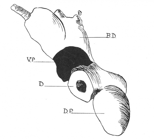
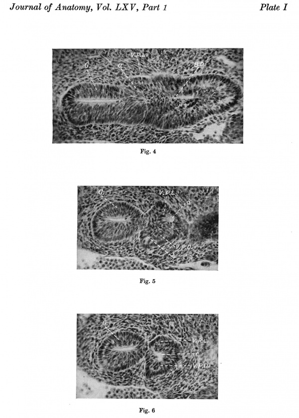
5mm.embryo. Here the ventral pancreas is seen filing up the angle that the hepatic outgrowth makes with the bowel and partially encircling the latter. It is horseshoe-shaped with a left limb smaller than the more projecting right limb, but commencing somewhat cranial to the latter. The whole bibbed outgrowth already appears as if it were being drawn out dorsalwards and to theright(fig.1). If sections (lOu) of this are examined, one finds that the ventral pancreas extends through seven sections in al. The left "lobe" appears first, two sections cranial to the right one, as a projection from the most caudal part of the opening of the bile duct into the gut (Plate I, fig. 4). If one looks at a section midway through this pancreas, one sees a definitely bilobed mass and in this, I suggest, are two lumina, of which the leftisthe larger and the more obvious (fig. 5). In the next section below this one (fig. 6) these two lumina are apparently fusing into one-a fine septum is seen dividing the common lumen-and the rest of the pancreas caudal to this is solid. Traced cranialwards, in the next section cranial to fig. 5 the right lumen has disappeared, but I can trace the left lumen through this one and the section next above it to open, I think, into the bile duct (fig. 4).
Fig. 1. Drawing of a model of the duodenum and pancreas, viewed from below, in a 5 mm. embryo. D. duodenum; B.D. bile-duct; V.P. ventral pancreas; D.P. dorsal pancreas.
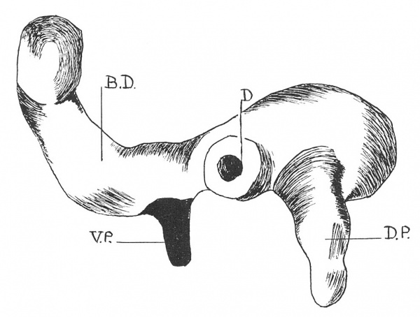
7.1 mm. embryo. The ventral pancreas here extends over three sections (10 micron) and is seen as a solid outgrowth from the right or dorsal side of the bile duct, which commences 70 micron cranial to the latter's opening into the gut (fig. 2). This outgrowth projects in a dorsal direction and is exactly parallel to the dorsal pancreas. The only suggestion of any lumen I can see in it is at its commencement, where the lumen of the bile duct appears to be slightly drawn out towards it in the form of a crescent. In neither the 5 mm. nor in the 7.1 mm. stage could I see any difference between the cells of the left or of the right portions of the outgrowths, suggesting that any of it was degenerating.
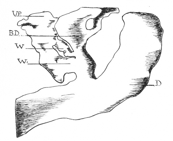
11.4 mm.embryo. Fig.3 shows a drawing of the ventral pancreas from a photograph of a reconstruction at this stage. In it one can see two ventral pancreatic ducts, one cranial to, and much smaller than the other: these run parallel to each other and both open, one immediately above its felow, into the common bileduct. I attempted to trace up these ducts to discover if they drain two distinct lobes of the ventral pancreas, but they both appear to issue from a fused mass of gland.
Fig. 2. Drawing of a model of the duodenum and pancreas, viewed from below, in a 7-1 mm. embryo. D. duodenum; B.D. bile-duct; V.P. ventral pancreas; D.P. dorsal pancreas.
These few observations of mine fal, I think, into line pretty well with those of previous observers. In the youngest, 5 mm. embryo, the ventral pancreas appears bibbed with two lumina: in the older, 7-1 mm. stage, there is but a single outgrowth, which appears to correspond to the right horn of the earlier one. This agrees entirely with Thyng's description of a slightly older 7-5 mm. embryo and with Lewis, who found a similar single outgrowth in the seven embryos of the Harvard Collection between 7.5 and 11 mm. in length. It is, however, hardly fair to assume from these later embryos, that the outgrowth was originally unpaired, and it is rather in the interpretation of the earlier stages that differences of opinion arise. As Siwe says, "In mehreren anderen FMillen kann man cine Andeutung zu Paarigkeit sehen- wenn man so will (oder auch nicht, wenn man nichtwill)." Iwould suggest that there are originally two separate outgrowths, right and left, which fuse very early. Of these the right is the more extensive. M. Tribe, in describing the development of the pancreas in marsupials, shows that the right ventral outgrowth contributes four times as much as does the left to the adult gland and remarks "in man the two ventral outgrowths are of such unequal size that one of them has frequently been overlooked altogether." This more extensive right lobed rag sits fellow to the right and dorsally.This appearance has been noted already as beginning in the 5 mm. stage and its persistence leads to the appearance of a single outgrowth, which, by the growth of the bile duct, is entirely divorced from the bowel wall. The lumina I describe in the 5 mm. stage are exactly similar to those figured by Jankelowitz in his 4.9 mm. embryo. While I realise that too much stress should not be laid upon them or upon their apparent fusion, Siwe, I think, rightly says "that one can hardly dismiss the question as Helly does, when he believes that two lumina can occur in any homogeneous anlage. Helly has not shown that the anlage was ever originally solid and further these lumina appear so early that one rather has reason to assume that no compact stage preceded them. The other possibility that a lumen, branched to right and left, could be the primary formation is at least as much justified." The duplication of the ventral duct, which I found in my 11.4 mm. embryo, is most readily explained by a delayed fusion of two separate outgrowths. I would suggest that the more slender and more cranial of the ducts represents the original left ventral proliferation, while the larger and more caudal of the two is formed by the larger right outgrowth.
Fig. 3. Drawing of a model of the duodenum and ventral pancreas, viewed from the right side, in an 11.4 mm. embryo. D. duodenum; B.D. bile-duct; V.P. ventral pancreas; W and W1 mark the two ventral pancreatic ducts.
Again, fusion of the two ventral elements seems a more probable ex- planation of the appearances noted than to assume that the left outgrowth atrophies. Debeyre in 1909 found the two ventral anlages in a 4*5mm. embryo, but, as he could of course only find a single outgrowth in the 12 mm. stage,assumed that the left proliferation early disappears. I have already pointed out that in Helly's 11mm. embryo the apparent degeneration of one of the ventral masses, abnormally unfused at this age and entirely separated from each other by the bile duct, cannot be admitted as good evidence of the normal atrophy of one outgrowth.
Further, a paired origin for the ventral pancreas in Man should be expected from the way in which this is developed in other animals. Inalvertebrates except Cyclostomes, it arises from the fusion of bilateral anlages (Siwe). Again, certain anomalous conditions in the adult are most easily explained in this way. Duplication of Wirsung's duct, which has been observed oc- casionally, would be the result of delayed fusion of the two separate outgrowths (Hasse), while the occurrence of an annular pancreas can be best ascribed to abnormally directed growth of the left ventral lobe,"when the left lobe tends to encircle the intestine instead of extending into the ventral mesentery " (Lewis).
Two other points arise out of an examination of the older 14 mm. and 23 mm. embryos. The time of fusion between the dorsal and the ventral pancreas must be variable. Keibel and Elze in the Normentafeln described them as uniting between the 12 and the 14 mm. stage. Inmy 14mm. embryo, while there is considerable interlocking, it is quite easy to pick out the two portions infact,they are more distinct than in the earlier,11-4mm.,one. Siwe mentions that fusion had not commenced in one embryo of 17 mm. Certainlyinmy 23 mm. specimen it was guesswork attempting to separate dorsal from ventral pancreas, which had completely joined together.
Lastly, an examination ofthe reconstructions from the 11-4 and the 14 mm. embryos suggests that the ventral pancreas must form much less of the fully developed gland than is often stated. I think it cannot be responsible really for more than the middle lateral part of its head, immediately about the terminal portion of Wirsung's duct.
Summary and Conclusions
Reconstruction models ofthe pancreas were made from human embryos 5, 7-1, 11-4, 14 and 23 mm. in length. In the 5mm. embryo the ventral pancreas appears as a bibbed mass, in which two apparent lumina can be distinguished: this is replaced in the 7-1mm. stage by a single outgrowth.These observations rather tend to support the view that the ventral pancreas in man is developed by two separate proliferations from the hepatic diverticulum, both of which, fusing early, may share in the formation of the adult gland. This view finds confirmation in the11-4mm.embryo examined. Here there is a duplication of the ventral duct, apparently due to delayed fusion of the two ventral anlages.
I should like to express my thanks to Prof. Arthur Thomson for much helpful advice and to Mr W. Chesterman for a great deal of assistance in preparing the reconstructions and for producing these microphotographs.
References
AREY, L. B. Developmental Anatomy.
BROMAN, I. Die Entwicklung des Menschen vor der Geburt.
DEBEYRE, M. A. (1909). C.R. de l'Assoc. des Anat.
FELIX,W. (1892). Arch. f. Anat. u. Phys.
HASSE, C. (1908). Anat. Anz. Bd. xxxii.
HELLY, K. (1901). Arch. f. Mikr. Anat. Bd. LVII.
INGGALLS, N. W. (1907). Arch. f. Mikr. Anat. Bd. LXX.
JANKELOWITZ, A. (1895). Arch. f. Mikr. Anat. Bd. XLVI.
KEIBEL and MALL. Manual of Human Embryology.
KEIBEL, F. and ELZE, C. (1908). Normentafeln.
KEITH, A. Human Embryology and Morphology.
LEWIS,F.T. (1911-12). Amer. J. of Anat. vol. XII.
MCMURRICHT, J.P. The Development of the Human Body.
OPIE, E.L. (1902). Bulletin of J. Hopkins Hospital.
PIPER, H. (1900). Arch. f. Anat. u. Entw.
SIWE, S. A. (1927). Jahrb. f. Morph. u. Mikr. Anat. Bd. LVII.
THYNG, F. W. (1907-8). Amer. J. of Anat. vol. VII.
TRIBE, M. (1918). Phil.Trans. B, vol. CCVIII.
ZIMMERMANN, K. W. (1898). Arch. f. Micr. Anat. Bd. LII.
Description of Plate 1
Fig.4. Section through the caudal end of the hepatic outgrowth. G. gut; B.D. bile-duct; V.P.L. left ventral pancreas, showing at a an apparent lumen opening into the junction of the bile-duct with the duodenum. (x200.)
Fig.5. Section midway through the ventral pancreas. G. gut; V.P.L. and V.P.D. mark the bilobed ventral pancreas, in which at a and bare two separate lumina. (x200.)
Fig. 6. Section immediately caudal to fig. 5. a. gut; V.P.L. and V.P.D. mark the ventral pancreas, in which atc there is a single lumen divided by a fine septum. (x200.)
PubMed Reference
Odgers PN. Some observations on the development of the ventral pancreas in man. (1930) J. Anat., 65(1): 1-7. PMID 17104298
Cite this page: Hill, M.A. (2024, April 26) Embryology Paper - Some Observations on the Development of the Ventral Pancreas in Man. Retrieved from https://embryology.med.unsw.edu.au/embryology/index.php/Paper_-_Some_Observations_on_the_Development_of_the_Ventral_Pancreas_in_Man
- © Dr Mark Hill 2024, UNSW Embryology ISBN: 978 0 7334 2609 4 - UNSW CRICOS Provider Code No. 00098G


