Movies - Chicken Neural Crest
Introduction
The images below link to pages containing each individual chicken neural crest movie, kindly provided by Paul Kulesa.[1]
In ovo time-lapse analysis of chick hindbrain neural crest cell migration shows cell interactions during migration to the branchial arches.[1]
- "Hindbrain neural crest cells were labeled with DiI and followed in ovo using a new approach for long-term time-lapse confocal microscopy. In ovo imaging allowed us to visualize neural crest cell migration 2-3 times longer than in whole embryo explant cultures, providing a more complete picture of the dynamics of cell migration from emergence at the dorsal midline to entry into the branchial arches." (More? full abstract)
Use the link to open the original movie in a new page. Each image in the movies represents 10 confocal sections separated by 10 microns each, projected onto 1 image.
| Neural crest migration Chicken Head (movies overview) | |||||||||||||||||||||||||||
|---|---|---|---|---|---|---|---|---|---|---|---|---|---|---|---|---|---|---|---|---|---|---|---|---|---|---|---|
|
|
|
|
|
|
| |||||||||||||||||||||


Interested in the cranial neural crest migration? See also mouse cranial neural crest.
About DiI
- a hydrophobic and lipophilic cyanine dye used for cell tracking as it is retained in the lipid bilayers
- chemical name is 1,1'-dioctadecyl-3,3,3'3'-tetramethylindocarbocyanine perchlorate
- the chemical formula is C59H89ClN2O4.
DiI-Labeled Neural Crest Cells

|
Chicken embryo sequence shows the migration of DiI-labeled neural crest cells towards the branchial arches as the embryo. White rings indicate migration of individual cells.
Legend
|

|
Chicken embryo sequence sequence shows the migration of DiI-labeled neural crest cells towards the branchial arches as the embryo undergoes its rotation to one side. Notice how the cells emigrate in streams which spread out to cover a subregion of the periphery.
Duration: 12 hrs. Time interval between images: 3 min. |
|
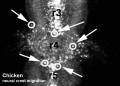
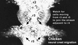
Chicken embryo sequence sequence shows the migration of DiI-labeled neural crest cells from r3, r4 and r5 contribute to the stream exiting adjacent to r4.
Duration: 3 hrs Time interval between images: 3 min |
|
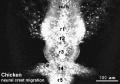
Chicken embryo sequence sequence shows the migration of DiI-labeled neural crest cells from r3 follow a caudolateral trajectory to join cells exiting adjacent to r4.
Duration: 4 hrs Time interval between images: 3 min |

|
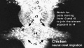
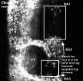
Chicken embryo sequence sequence shows the migration of DiI-labeled neural crest cells move into the regions between the streams and make contact with cells from a different stream. In this movie, neural crest cells from the first branchial arch stream migrate caudally towards the second branchial arch. And neural crest cells at the caudal part of the second branchial arch stream meet with cells from the third branchial arch stream.
Duration: 2 hrs Time interval between images: 3 min |
|
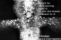
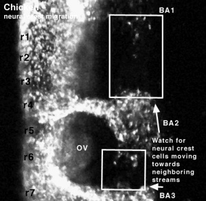
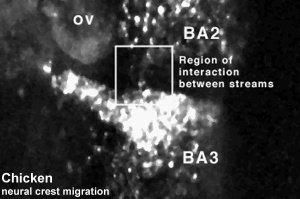
Chicken embryo sequence sequence shows the migration of DiI-labeled neural crest cells zoomed in on the region of neural crest cell interactions lateral to the otic vesicle. This sequence of images shows neural crest cells from the second and third branchial arch streams interacting.
Duration: 3 hrs Time interval between images: 3 min |
|
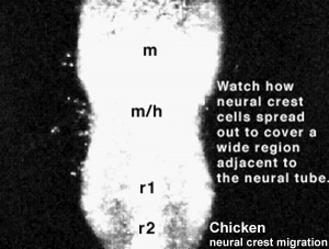
Chicken embryo sequence sequence shows the migration of DiI-labeled neural crest cells leaving from near the midbrain (m), midbrain/hindbrain boundary (m/h) and rostral rhombomeres (r1 and r2) and spread out to cover a wide region adjacent to the neural tube.
Duration: 7 hrs Time interval between images: 3 min |
m = midbrain h = hindbrain r = rhombomere
Each image in the movies represents 10 confocal sections separated by 10 microns each, projected onto 1 image.
Movies Source: Original Neural Crest movies kindly provided by Paul Kulesa.
Neural Crest Movie Links: Movie 1 | Neural Crest Development | Chicken Development | Movies
Links: Movies - Chicken Neural Crest | Neural Crest Development | all Development movies
References
Glossary Links
- Glossary: A | B | C | D | E | F | G | H | I | J | K | L | M | N | O | P | Q | R | S | T | U | V | W | X | Y | Z | Numbers | Symbols | Term Link
Cite this page: Hill, M.A. (2024, April 26) Embryology Movies - Chicken Neural Crest. Retrieved from https://embryology.med.unsw.edu.au/embryology/index.php/Movies_-_Chicken_Neural_Crest
- © Dr Mark Hill 2024, UNSW Embryology ISBN: 978 0 7334 2609 4 - UNSW CRICOS Provider Code No. 00098G