Lymph Node Development: Difference between revisions
| Line 80: | Line 80: | ||
</gallery> | </gallery> | ||
==Cell Trafficking into and out of Lymph Nodes== | |||
[[File:Lymph_node_cartoon_02.jpg|800px]] | [[File:Lymph_node_cartoon_02.jpg|800px]] | ||
Revision as of 10:48, 28 February 2012
Introduction


Lymphatic vasculature drains lymph fluid from the organ tissue space and returns it to the blood vasculature for recirculation. Lymph nodes lie on the path of lymph vessels and these structures monitor and carry out immune surveillance of this fluid for antigens and pathogens, trapping them within the lymph nodes and generating immune responses.
Some Recent Findings
|
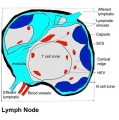
Adult Lymph Node
- Encapsulated organ (1 mm - 2 cm)
- In lymph vessel pathways “filter”
- Afferent- towards node
- Efferent- away from node
- Location throughout the entire body - Concentrated in axilla, groin, mesenteries
- Antigen transformed lymphocytes from the blood
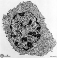
Lymph Node Cartoon Gallery
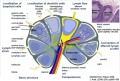
Detailed structure
Detailed structure
Simple structure
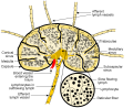
Wiki image
internal structure


- Links: Immunobiology - Figure 1.8. Organization of a lymph node | MBoC Figure 24-16. A simplified drawing of a human lymph node

|
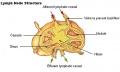
Schematic representation of the organization of a lymph node.[2]
|

Histology
- Lymph Node Histology: Subcapsular Sinus | Follicle | Germinal Centre | Medullary Cords and Sinuses | High Endothelial Venules | Macrophages | Node cartoons
Adult Lymph Node Structure
- Capsule - dense connective tissue
- Trabeculae - dense connective tissue
- Reticular Tissue - Reticular cells and fibers, supporting meshwork
- Macrophages - process antigen, difficult to distinguish from the reticular cells.


Lymph
- enters the node through afferent vessels
- filters through the sinuses
- leaves through efferent vessels
Subcapsular sinus = marginal sinus

Continuation of trabecular sinus

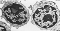
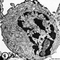
Adult Lymphocytes
Lymphocyte Electron Micrographs
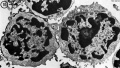
T and B Lymphocyte
T and B Lymphocyte
Plasma cell (B)
Cytotoxic (T)


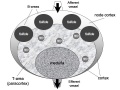
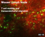
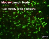
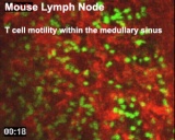
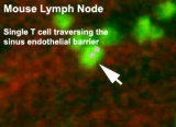
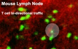
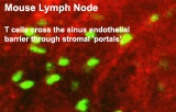
Cell Trafficking into and out of Lymph Nodes

Lymphocyte Traffic in and out of the Lymph Node[3]
The following data is from a recent article[4] and review[5] of live adult mouse lymphocytes (T and B cells) imaged within a lymph node.
Both lymphocyte types:
- Spend 8 to 24 h in the lymph node interstitium.
- Transit across a lymphatic endothelium to exit.
- Enter a network of medullary sinuses.
- Drain from sinuses into efferent lymphatic vessels.
Lymphocyte Migration Speeds
T cells - 10–12 μm/min in the follicle diffuse cortex, peak velocities up to 30 μm/min. (move more slowly in the medullary region near the hilus of the lymph node than in the paracortex)
B cells - 6 μm/min in the follicle diffuse cortex, peak velocities up to 20 μm/min.
Both cortical T cells and follicular B cells move in random directions following "guide cells".
Lymphocyte Guide Cells
FDC - Follicular Dendritic Cells, may guide B cells in the follicle.
FRC - Fibroblastic Reticular Cells, may guide T cells in the follicle.
Lymphocyte Movies
Adult Mouse Lymph Node - T cell motility
|
|
|
| Transendothelial migration | T cell zone | Medullary sinus |
|
|
|
| Sinus endothelial barrier | Bi-directional traffic | Cross the sinus endothelial barrier |
References
- ↑ <pubmed>19060331</pubmed>
- ↑ <pubmed>19644499</pubmed>| PMC2785037 | Nat Rev Immunol.
- ↑ <pubmed>15122201</pubmed>| Nat Rev Immunol.
- ↑ <pubmed>16273098</pubmed>
- ↑ <pubmed>18173372</pubmed>
Textbook
Immunobiology 5th edition The Immune System in Health and Disease Charles A Janeway, Jr, Paul Travers, Mark Walport, and Mark J Shlomchik.
Part I. An Introduction to Immunobiology and Innate Immunity
- Chapter 1. Basic Concepts in Immunology
- The components of the immune system
- Figure 1.3 All the cellular elements of blood, including the lymphocytes of the adaptive immune system, arise from hematopoietic stem cells in the bone marrow
- Figure 1.4 Myeloid cells in innate and adaptive immunity
- Figure 1.5 Lymphocytes are mostly small and inactive cells
- Figure 1.6 Natural killer (NK) cells
- Figure 1.7 The distribution of lymphoid tissues in the body
- Figure 1.8 Organization of a lymph node
- Figure 1.9 Organization of the lymphoid tissues of the spleen
- Summary to Chapter 1
- The components of the immune system
Part III. The Development of Mature Lymphocyte Receptor Repertoires
- Chapter 7. The Development and Survival of Lymphocytes
Reviews
<pubmed></pubmed>
Articles
<pubmed>165702</pubmed> <pubmed>1167215</pubmed>
Search Pubmed
Search Pubmed: Lymph Node Development | Lymphocyte Development
Glossary Links
- Glossary: A | B | C | D | E | F | G | H | I | J | K | L | M | N | O | P | Q | R | S | T | U | V | W | X | Y | Z | Numbers | Symbols | Term Link
Cite this page: Hill, M.A. (2024, May 4) Embryology Lymph Node Development. Retrieved from https://embryology.med.unsw.edu.au/embryology/index.php/Lymph_Node_Development
- © Dr Mark Hill 2024, UNSW Embryology ISBN: 978 0 7334 2609 4 - UNSW CRICOS Provider Code No. 00098G