Human Sylvian Fissure Movie: Difference between revisions
No edit summary |
No edit summary |
||
| Line 1: | Line 1: | ||
{{Movie header}} | {{Movie header}} | ||
{| | {| | ||
| width=360px|<mediaplayer width='355' height='390' image="http://embryology.med.unsw.edu.au/embryology/images/e/e6/Brain_fissure_development_03.jpg">File:Neural_-_Sylvian_fissure.mp4</mediaplayer> | | width=360px|<mediaplayer width='355' height='390' image="http://embryology.med.unsw.edu.au/embryology/images/e/e6/Brain_fissure_development_03.jpg">File:Neural_-_Sylvian_fissure.mp4 </mediaplayer> | ||
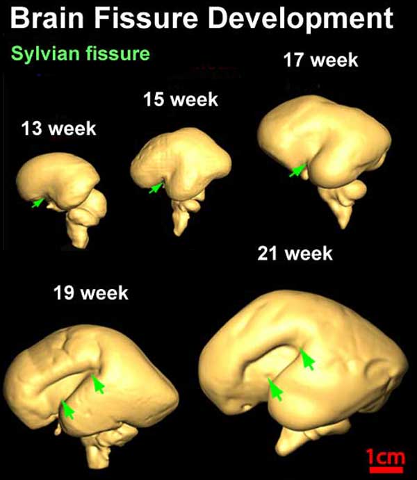
| valign="top" | This is an animation created from individual three-dimensional reconstruction of the lateral (top row) surface of 13–21 week brains to reveal the development of the Sylvian or lateral fissure (green arrow). | | valign="top" | This is an animation created from individual three-dimensional reconstruction of the lateral (top row) surface of 13–21 week brains to reveal the development of the Sylvian or lateral fissure (green arrow). | ||
Revision as of 12:53, 8 March 2013
| Embryology - 26 Apr 2024 |
|---|
| Google Translate - select your language from the list shown below (this will open a new external page) |
|
العربية | català | 中文 | 中國傳統的 | français | Deutsche | עִברִית | हिंदी | bahasa Indonesia | italiano | 日本語 | 한국어 | မြန်မာ | Pilipino | Polskie | português | ਪੰਜਾਬੀ ਦੇ | Română | русский | Español | Swahili | Svensk | ไทย | Türkçe | اردو | ייִדיש | Tiếng Việt These external translations are automated and may not be accurate. (More? About Translations) |
| <mediaplayer width='355' height='390' image="http://embryology.med.unsw.edu.au/embryology/images/e/e6/Brain_fissure_development_03.jpg">File:Neural_-_Sylvian_fissure.mp4 </mediaplayer> | This is an animation created from individual three-dimensional reconstruction of the lateral (top row) surface of 13–21 week brains to reveal the development of the Sylvian or lateral fissure (green arrow).
|

Diffusion tensor imaging (DTI) A newly developed form of magnetic resonance imaging (MRI). Magnetic field variations of the MRI magnet are applied in at least six different directions generating a three dimensional shape of the diffusion pattern. This technique can be used in neural imaging of white matter due to the orientation of axon bundles and the associated water flow. (More? Magnetic Resonance Imaging)
- Neural DTI Links: Scaled Fissures 13-21 weeks | Fissures 13-21 weeks | Brain Sylvian Fissure | Scaled Brain and Ventricles 13-21 weeks | Scaled Brain, Ventricles and Ganglia 13-21 weeks | Limbic Tract 13-19 weeks | Brain and Ventricles 13-21 weeks | Sylvian Fissure Movie | Neural System Development | Magnetic Resonance Imaging
Reference
<pubmed>19339620</pubmed>| PMC2721010 | J Neurosci.
Original File Name: Figure 5 Original image modified by scaling relative to 21 weeks, labeling, and reorganizing arrangement of image.
Glossary Links: A | B | C | D | E | F | G | H | I | J | K | L | M | N | O | P | Q | R | S | T | U | V | W | X | Y | Z | Numbers | Symbols | Movies
Cite this page: Hill, M.A. (2024, April 26) Embryology Human Sylvian Fissure Movie. Retrieved from https://embryology.med.unsw.edu.au/embryology/index.php/Human_Sylvian_Fissure_Movie
- © Dr Mark Hill 2024, UNSW Embryology ISBN: 978 0 7334 2609 4 - UNSW CRICOS Provider Code No. 00098G