HM Practical - Cardiac Histology: Difference between revisions
From Embryology
| Line 37: | Line 37: | ||
* At high magnification see both striations and the large nuclei of the cardiac muscle cells. | * At high magnification see both striations and the large nuclei of the cardiac muscle cells. | ||
* Follow the course of individual cardiac muscle cells and note fine, dark blue lines which seem to cross (traverse) the fibres. | * Follow the course of individual cardiac muscle cells and note fine, dark blue lines which seem to cross (traverse) the fibres. | ||
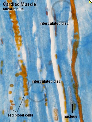
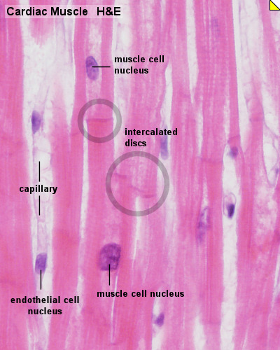
* | * '''Intercalated Discs''' that connect the individual muscle cells and permit the conduction of electrical impulses between the cells. | ||
** seen in longitudinal sections. | |||
|} | |} | ||
| Line 54: | Line 56: | ||
File:Heart_histology_107.jpg | File:Heart_histology_107.jpg | ||
</gallery> | </gallery> | ||
===Intercalated Discs=== | |||
* seen in longitudinal sections. | |||
* connect the individual muscle cells. | |||
* permit the conduction of electrical impulses between the cells. | |||
===Purkinje Fibres=== | ===Purkinje Fibres=== | ||
Revision as of 08:18, 6 August 2012
Introduction
HMA Practical 8 Monday August 6 and Wednesday August 8.
HMA Practical 8 Virtual Slides
This page provides histology support information for cardiac histology.
Disclaimers
- does not form part of the actual practical class based upon the virtual slides.
- does not cover the pathology content.
- HMA Links: Blood Vessel Histology | Cardiac Histology | Histology | Histology Stains | Blood Vessel Development | HMA Practical 3 Virtual Slides | HMA Practical 8 Virtual Slides
Introduction
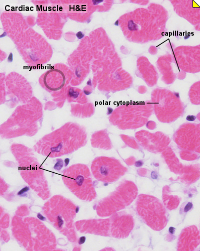
Cardiac muscle, the myocardium, consists of cross-striated muscle cells, cardiomyocytes, with one centrally placed nucleus.
- Nuclei are oval, rather pale and located centrally in the muscle cell which is 10 - 15 µm wide.
- Cardiac muscle cells excitation is mediated by rythmically active modified cardiac muscle cells.
- Cardiac muscle is innervated by the autonomic nervous system (involuntary), which adjusts the force generated by the muscle cells and the frequency of the heart beat.
- Cardiac muscle cells often branch at acute angles and are connected to each other by specialisations of the cell membrane in the region of the intercalated discs.
- Intercalated discs invariably occur at the ends of cardiac muscle cells in a region corresponding to the Z-line of the myofibrils.
- Cardiac muscle does not contain cells equivalent to the satellite cells of skeletal muscle.
Histology
Unlabeled Images
Intercalated Discs
- seen in longitudinal sections.
- connect the individual muscle cells.
- permit the conduction of electrical impulses between the cells.
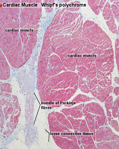
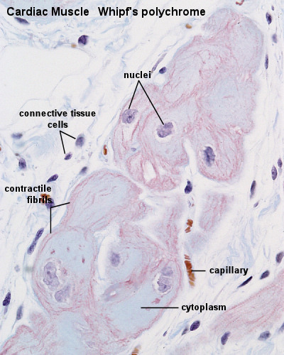
Purkinje Fibres
- modified cardiac muscle cells. Compared to ordinary cardiac muscle cells:
- contain large amounts of glycogen.
- fewer myofibrils.
- thicker cells.
- extend from the atrioventricular node, pierces the fibrous body, divides into left and right bundles, and travels, beneath the endocardium, towards the apex of the heart.
- bundle branches contact cardiac muscle cells through specialisations similar to intercalated discs.
- conduct stimuli faster than ordinary cardiac muscle cells (2-3 m/s vs. 0.6 m/s).
- discovered in 1839 by Jan Evangelista Purkyně).
- Links: Heart Histology | Cardiac AZB Labeled | Cardiac AZB | Cardiac label LS | Cardiac LS | Cardiac label TS | Cardiac TS | Purkinje fibres | Purkinje fibres detail | Histology
Terms
- cardiomyocyte -
- intercalated disc -
- Purkinje fibres -
Glossary Links
- Glossary: A | B | C | D | E | F | G | H | I | J | K | L | M | N | O | P | Q | R | S | T | U | V | W | X | Y | Z | Numbers | Symbols | Term Link
Cite this page: Hill, M.A. (2024, May 30) Embryology HM Practical - Cardiac Histology. Retrieved from https://embryology.med.unsw.edu.au/embryology/index.php/HM_Practical_-_Cardiac_Histology
- © Dr Mark Hill 2024, UNSW Embryology ISBN: 978 0 7334 2609 4 - UNSW CRICOS Provider Code No. 00098G
