File:Thyroid pyramidal lobe.jpg
Thyroid_pyramidal_lobe.jpg (300 × 338 pixels, file size: 19 KB, MIME type: image/jpeg)
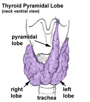
Thyroid Abnormality - Pyramidal Lobe
A common thyroid anatomical variation seen in as high as half anatomical dissections and more frequently in men than in women.[1] Because of the common nature of this anatomical variation and often with no impact upon thyroid function, it may not be classified as a true developmental abnormality.
A third lobe, of conical shape, called the pyramidal lobe, frequently arises from the upper part of the isthmus, or from the adjacent portion of either lobe, but most commonly the left, and ascends as far as the hyoid bone. It is occasionally quite detached, or may be divided into two or more parts.
- Links: thyroid
References
Cite this page: Hill, M.A. (2024, April 27) Embryology Thyroid pyramidal lobe.jpg. Retrieved from https://embryology.med.unsw.edu.au/embryology/index.php/File:Thyroid_pyramidal_lobe.jpg
- © Dr Mark Hill 2024, UNSW Embryology ISBN: 978 0 7334 2609 4 - UNSW CRICOS Provider Code No. 00098G
File history
Click on a date/time to view the file as it appeared at that time.
| Date/Time | Thumbnail | Dimensions | User | Comment | |
|---|---|---|---|---|---|
| current | 10:46, 6 October 2009 | | 300 × 338 (19 KB) | S8600021 (talk | contribs) | Thyroid abnormalities - pyramidal lobe Category:Endocrine Category:Thyroid Category:Abnormal Development |
You cannot overwrite this file.