File:Streeter1919-fig01.jpg: Difference between revisions
mNo edit summary |
mNo edit summary |
||
| (2 intermediate revisions by the same user not shown) | |||
| Line 1: | Line 1: | ||
==Fig. 1. Profile reconstructions showing the spinal ganglia and their dorsal roots in the tail region of the human embryo== | ==Fig. 1. Profile reconstructions showing the spinal ganglia and their dorsal roots in the tail region of the human embryo== | ||
white - Last two lumbar ganglia | |||
stippled - Sacral ganglia | |||
solid black - coccygeal ganglia | |||
It will be noted that in the period included between these two stages marked regressive changes have aflected the entire coccygeal region of the spinal cord, with complete disappearance of the last three coccygeal ganglia, in sharp contrast to the sacral region of the cord, which undergoes uninterrupted development. The reconstructions are taken from embryos No. {{CE544}}, 11.5 mm, long, and No. {{CE75}}, 30 mm. long, belonging to the [[Carnegie Collection]]. | |||
{{Streeter1919 figures}} | {{Streeter1919 figures}} | ||
[[Category:Carnegie Embryo 544]][[Category:Carnegie Embryo 75]] | |||
Latest revision as of 14:07, 25 May 2018
Fig. 1. Profile reconstructions showing the spinal ganglia and their dorsal roots in the tail region of the human embryo
white - Last two lumbar ganglia
stippled - Sacral ganglia
solid black - coccygeal ganglia
It will be noted that in the period included between these two stages marked regressive changes have aflected the entire coccygeal region of the spinal cord, with complete disappearance of the last three coccygeal ganglia, in sharp contrast to the sacral region of the cord, which undergoes uninterrupted development. The reconstructions are taken from embryos No. 544, 11.5 mm, long, and No. 75, 30 mm. long, belonging to the Carnegie Collection.
| Historic Disclaimer - information about historic embryology pages |
|---|
 |
Reference
Streeter GL. Factors involved in the formation of the filum terminale. (1919) Amer. J Anat. 22(1): 1-11.
Cite this page: Hill, M.A. (2024, May 18) Embryology Streeter1919-fig01.jpg. Retrieved from https://embryology.med.unsw.edu.au/embryology/index.php/File:Streeter1919-fig01.jpg
- © Dr Mark Hill 2024, UNSW Embryology ISBN: 978 0 7334 2609 4 - UNSW CRICOS Provider Code No. 00098G
File history
Click on a date/time to view the file as it appeared at that time.
| Date/Time | Thumbnail | Dimensions | User | Comment | |
|---|---|---|---|---|---|
| current | 23:53, 11 September 2015 | 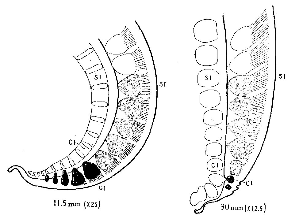 | 975 × 716 (126 KB) | Z8600021 (talk | contribs) | |
| 23:44, 11 September 2015 |  | 1,355 × 1,149 (301 KB) | Z8600021 (talk | contribs) |
You cannot overwrite this file.
File usage
The following 3 pages use this file: