File:Stage8 bf4.jpg
Stage8_bf4.jpg (600 × 449 pixels, file size: 17 KB, MIME type: image/jpeg)
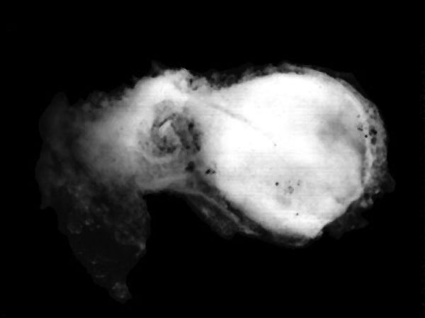
Human Embryo
Features: embryonic disc, primitive node, primative streak, primative groove, connecting stalk
Facts: Week 3, 17 - 19 days, 1.0 - 1.5 mm
View: embryonic disc, showing the epiblast viewed from the amniotic (dorsal) side. Amniotic membrane removed, connecting stalk to the left.
Events: Gastrulation is continuing as cells migrate from the epiblast, continuing to form mesoderm.
Mesoderm lies between the ectoderm and endoderm as a continuous sheet except at the buccopharyngeal and cloacal membranes. These membranes have ectoderm and endoderm only and will lie at the rostral (head) and caudal (tail) of the gastrointestinal tract.
From the primitive node a tube extends under the ectoderm in the opposite direction to the primitive streak. This tube forms first the axial process then notochordal process, then finally the notochord.
The notochord is a key to embryonic folding and regulation of ectoderm and mesoderm differentiation. It lies in the rostrocordal axis and the embryonic disc will fold either side ventrally, pinching off a portion of the yolk sac to form the lining of the gastrointestinal tract.
Original file name: http://embryology.med.unsw.edu.au/wwwhuman/Stages/Images/CSt8.gif
Image source: The Kyoto Collection images are reproduced with the permission of Prof. Kohei Shiota and Prof. Shigehito Yamada, Anatomy and Developmental Biology, Kyoto University Graduate School of Medicine, Kyoto, Japan for educational purposes only and cannot be reproduced electronically or in writing without permission.
File history
Click on a date/time to view the file as it appeared at that time.
| Date/Time | Thumbnail | Dimensions | User | Comment | |
|---|---|---|---|---|---|
| current | 20:24, 15 March 2011 | | 600 × 449 (17 KB) | S8600021 (talk | contribs) | == Human Embryo == Features: embryonic disc, primitive node, primative streak, primative groove, connecting stalk Facts: Week 3, 17 - 19 days, 1.0 - 1.5 mm View: embryonic disc, showing the epiblast viewed from the amniotic (dorsal) side. Amniotic memb |
You cannot overwrite this file.
File usage
The following 14 pages use this file:
- Carnegie Stages
- Carnegie stage 8
- Carnegie stage table
- Embryonic Development
- Kyoto Collection
- Lecture - Mesoderm Development
- Paper - A Human Embryo of Twenty-five Somites
- Paper - A Human Embryo of Twenty-seven Pairs of Somites, Embedded in Decidua
- Paper - Report upon the collection of human embryos at the Johns Hopkins University (1911)
- Paper - Two presomite human embryos
- Talk:Carnegie Stages
- Template:Carnegie stage table
- Template talk:Carnegie stage table
- History:Paper - A Human Embryo of Twenty-seven Pairs of Somites, Embedded in Decidua