File:Spinal cord histology 09.jpg
Original file (1,280 × 1,024 pixels, file size: 227 KB, MIME type: image/jpeg)
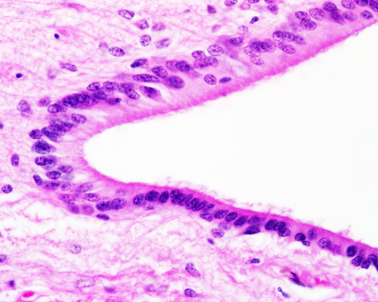
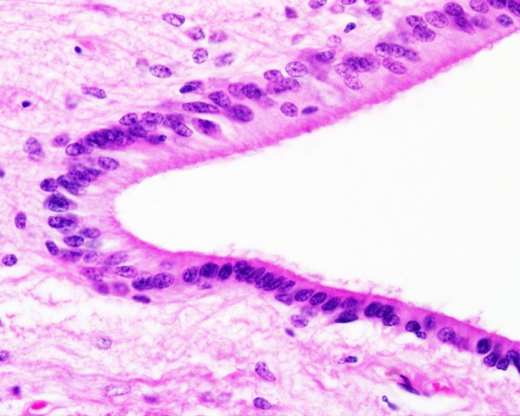
central canal, ependymal cells
- Tissue - sheep spinal cord
- Stain - H&E
- Spinal Cord: Overview 1 | Overview 2 | Overview animation | Grey matter | Grey matter | Grey matter | White matter | Overview unlabeled | Grey matter unlabeled 1 | Grey matter unlabeled 2 | White matter unlabeled 1 | Ependymal cells unlabeled
Links: Histology | Histology Stains | Blue Histology images copyright Lutz Slomianka 1998-2009. The literary and artistic works on the original Blue Histology website may be reproduced, adapted, published and distributed for non-commercial purposes. See also the page Histology Stains.
Cite this page: Hill, M.A. (2024, April 27) Embryology Spinal cord histology 09.jpg. Retrieved from https://embryology.med.unsw.edu.au/embryology/index.php/File:Spinal_cord_histology_09.jpg
- © Dr Mark Hill 2024, UNSW Embryology ISBN: 978 0 7334 2609 4 - UNSW CRICOS Provider Code No. 00098G
Spinal cord histology 09.jpg Original file name: epen40he.jpg
File history
Click on a date/time to view the file as it appeared at that time.
| Date/Time | Thumbnail | Dimensions | User | Comment | |
|---|---|---|---|---|---|
| current | 10:32, 20 September 2012 | | 1,280 × 1,024 (227 KB) | Z8600021 (talk | contribs) |
You cannot overwrite this file.
File usage
The following 4 pages use this file: