File:Sgalitzer1941 fig01.jpg

Original file (577 × 1,200 pixels, file size: 55 KB, MIME type: image/jpeg)
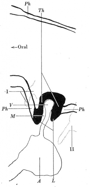
Fig 1 Mid-sagittal graphic reconstruction of the thyroid anlage of embryo F
The cavities of the first and second pharyngeal pouches of the right side have been projected on to the mid-sagital plane.
'A'Bold text, aortic sac; L, lipped margin of thyroid anlage; M, connective tissue core; Ph, pharyngeal epithelium; Th, thyroid anlage; F, its vessel; I, II, first and second pharyngeal pouches. x250.
Fig. 1 shows the mid-sagittal reconstruction of a part of the pharynx of embryo F (4 mm, 28 somites). The cavities of the first and second pharyngeal pouches of the right side have been projected on to the midsagittal plane in order to show that the thyroid anlage is situated at its normal level. The latter, however, instead of showing the normal evagination, is represented by a small nodule which projects into the pharyngeal cavity. It is formed by an epithelium which comparison shows to be similar in appearance to a normal thyroid primordium (fig. 2a). There projects into the nodule a mesodermal core containing a small vessel which arises from the aortic sac. The epithelial margins of the nodule are projected like lips towards the underlying connective tissue, and the outer part of these lips merges into the pharyngeal epithelium.
Reference
Sgalitzer KE. Contribution to the study of the morphogenesis of the thyroid gland. (1941) J Anat. 75(4): 389-405. PMID 17104869
Cite this page: Hill, M.A. (2024, April 27) Embryology Sgalitzer1941 fig01.jpg. Retrieved from https://embryology.med.unsw.edu.au/embryology/index.php/File:Sgalitzer1941_fig01.jpg
- © Dr Mark Hill 2024, UNSW Embryology ISBN: 978 0 7334 2609 4 - UNSW CRICOS Provider Code No. 00098G
File history
Click on a date/time to view the file as it appeared at that time.
| Date/Time | Thumbnail | Dimensions | User | Comment | |
|---|---|---|---|---|---|
| current | 14:13, 19 March 2017 | | 577 × 1,200 (55 KB) | Z8600021 (talk | contribs) | |
| 14:12, 19 March 2017 | 616 × 1,728 (136 KB) | Z8600021 (talk | contribs) | ===Reference=== {{Ref-Sgalitzer1941}} |
You cannot overwrite this file.
File usage
The following 2 pages use this file: