File:Sensenig1951 plate04.jpg

Original file (1,990 × 2,627 pixels, file size: 1.29 MB, MIME type: image/jpeg)
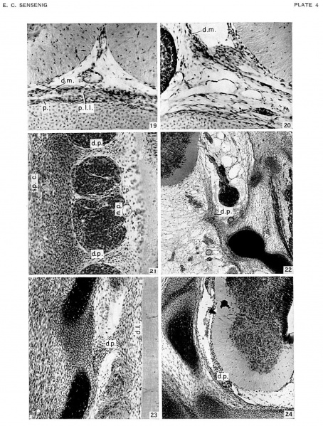
Plate 4
Fig. 19. Transverse section of lower cervical region of embryo no. 4473, 43.0 mm.. cut 20 microns. X230. Shows single cell layer of pia mater and nntcrior spinal zirlcry. also duru mnlcr and pcrichunilrium with rudimcm of puslcrior im1_=_:ituL|in;I| li;_v:nncnL i1L‘[\\'CL‘l1 thcm.
Fig. 20. Transverse section of upper thoracic region of embryo no. 4475. 48.0 mm. cut 20 microns x250. l’i:1 mater and durzl mnlcr lying mcdiui to spinal g:n1_-,;‘|iun.
Fig. 21. Frontal section of thoracic region of embryo no. 8113. 12.6 mm.. age group xvii, cut 10 microns. x200. Interganglionic tissue connecting dense laterally situated mesenchyme with lateral border of neural tube representing the earliest indication of a dentate process.
Fig. 22. Transverse section of occipitoccr\'ic:Il rc;.;run of embryo no. 7592. 32.7 mm.. age group xxi. cut :4» microns. Ex’ -‘,0. First dcnlulc proccss c.\‘1cndin;_: dorm-incdiml from rudiment of occipital bulk’ to ncurnl lnhc.
Fig. 23;. Frontal suction of mid-thoracic region of embryo no. 6832; 23.3 mm.. age group xxii. cut . 20 microns. x150. Dentate process joins latcralliy Ihc cerebral arch rudiment. and mcdislily u thickcnin_-.3 of the pininatcr. lhc rndimcnlnry dcnticulznc ligmncnl.
Fig. 24. Transverse section of upper thoracic region of embryo no. 4570, 30.7 mm.. age group xxiii. cut 15 microns.x100.Interganglionic section showing dentate process.
Abbreviations used in Plates: d.1., dentate ligament d.m., dura mater d.p., dentate process m.p., meninx primitiva n.c., neural crest n.c.c., neural-crest cells n.p., neural process p., perichondrium pm., rudimentary posterior longitudinal ligament s., somite :p.c., spinal cord :p.g., spinal ganglion
| Historic Disclaimer - information about historic embryology pages |
|---|
 |
- Links: plate 1 | plate 2 | plate 3 | plate 4 | Sensenig 1951 | Spinal Cord | Meninges
Reference
Sensenig EC. The early development of the meninges of the spinal cord in human embryos. (1951) Contrib. Embryol., Carnegie Inst. Wash. Publ. 611.
Cite this page: Hill, M.A. (2024, May 6) Embryology Sensenig1951 plate04.jpg. Retrieved from https://embryology.med.unsw.edu.au/embryology/index.php/File:Sensenig1951_plate04.jpg
- © Dr Mark Hill 2024, UNSW Embryology ISBN: 978 0 7334 2609 4 - UNSW CRICOS Provider Code No. 00098G
File history
Click on a date/time to view the file as it appeared at that time.
| Date/Time | Thumbnail | Dimensions | User | Comment | |
|---|---|---|---|---|---|
| current | 15:25, 5 June 2016 | | 1,990 × 2,627 (1.29 MB) | Z8600021 (talk | contribs) | ==Plate 4== {{Sensenig1951 images}} |
You cannot overwrite this file.
File usage
The following 2 pages use this file: