File:Pohlman1911 plate3F.jpg
Original file (1,418 × 1,512 pixels, file size: 98 KB, MIME type: image/jpeg)
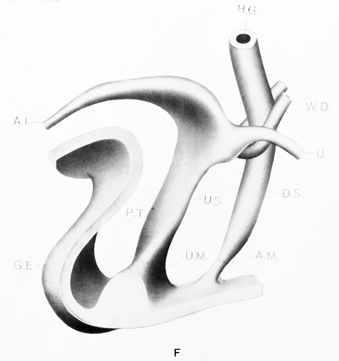
Plate 3F
Model F. {No. 24; Mall no. 43; 16.0 mm. X 50.) Unfortunately this embryo was cut too thick for minute reconstruction. The stage however fills in another gap in the Keibel series. Here the cloaca and the cloacal membrane are completely divided - the ventral segment limited by the urogenital plate and the dorsal segment by the anal plate. The ureter is displaced from its dorso-lateral position on the Wolffian duct to a supero-lateral one and opens distinctly into the ventral cloacal segment. The marked increase in precloacal tissue has resulted not only in the large genital eminence, but the urogenital plate has been dislocated deeper into the base of the phallus and a marked heaping up of ectodermal cells has occurred in the furrow on the caudal surface of the eminence. The two resultant segments of the cloacal membrane, the urogenital and anal plates are apparently no longer than they were in much younger stages; a point that will be brought out in greater detail later.
No. 24 (Mall 43; 16.0mm.; = ; 50; Good). Model F . x 50. Anal and urogenital membranes separated. Genital eminence more marked. Ectodermic inclusion in furrow on caudal surface. Ureteral orifice on same level but some distance lateral to Wolffian orifice. Carnegie stage 19
| Key to Lettering | ||
|---|---|---|
|
A., Allantois K., Kidney A.M., Position of anal membrane P.T., Precloacal mesodermic tissue C., Cloaca |
R.B., Renal bud CM., Cloacal membrane T.G., Tail cut C.S., Cloacal segment of Wolffian duct U., Ureter |
G.E., Genital eminence U.G., Urogenital sinus H.G., Hind gut U.M., Position of urogenital membrane * Probable position of point where allantois joins cloaca. |
All drawings represent 100 diameters enlargement except of Model F which is 50 diameters.
- Links: plate 1 | 1 A | 1 B | plate 2 | plate 2C | plate 2D | plate 3 | plate 3E | plate 3F | Pohlman 1911
Reference
Pohlman AG. The development of the cloaca in human embryos. (1911) Amer. J Anat. 12: 1-26.
Cite this page: Hill, M.A. (2024, April 27) Embryology Pohlman1911 plate3F.jpg. Retrieved from https://embryology.med.unsw.edu.au/embryology/index.php/File:Pohlman1911_plate3F.jpg
- © Dr Mark Hill 2024, UNSW Embryology ISBN: 978 0 7334 2609 4 - UNSW CRICOS Provider Code No. 00098G
File history
Click on a date/time to view the file as it appeared at that time.
| Date/Time | Thumbnail | Dimensions | User | Comment | |
|---|---|---|---|---|---|
| current | 14:55, 15 June 2016 | | 1,418 × 1,512 (98 KB) | Z8600021 (talk | contribs) | ==Plate 3F== {{Pohlman1911 figures}} |
You cannot overwrite this file.
File usage
The following 2 pages use this file: