File:Pohlman1911 plate1B.jpg
Original file (1,226 × 1,449 pixels, file size: 180 KB, MIME type: image/jpeg)
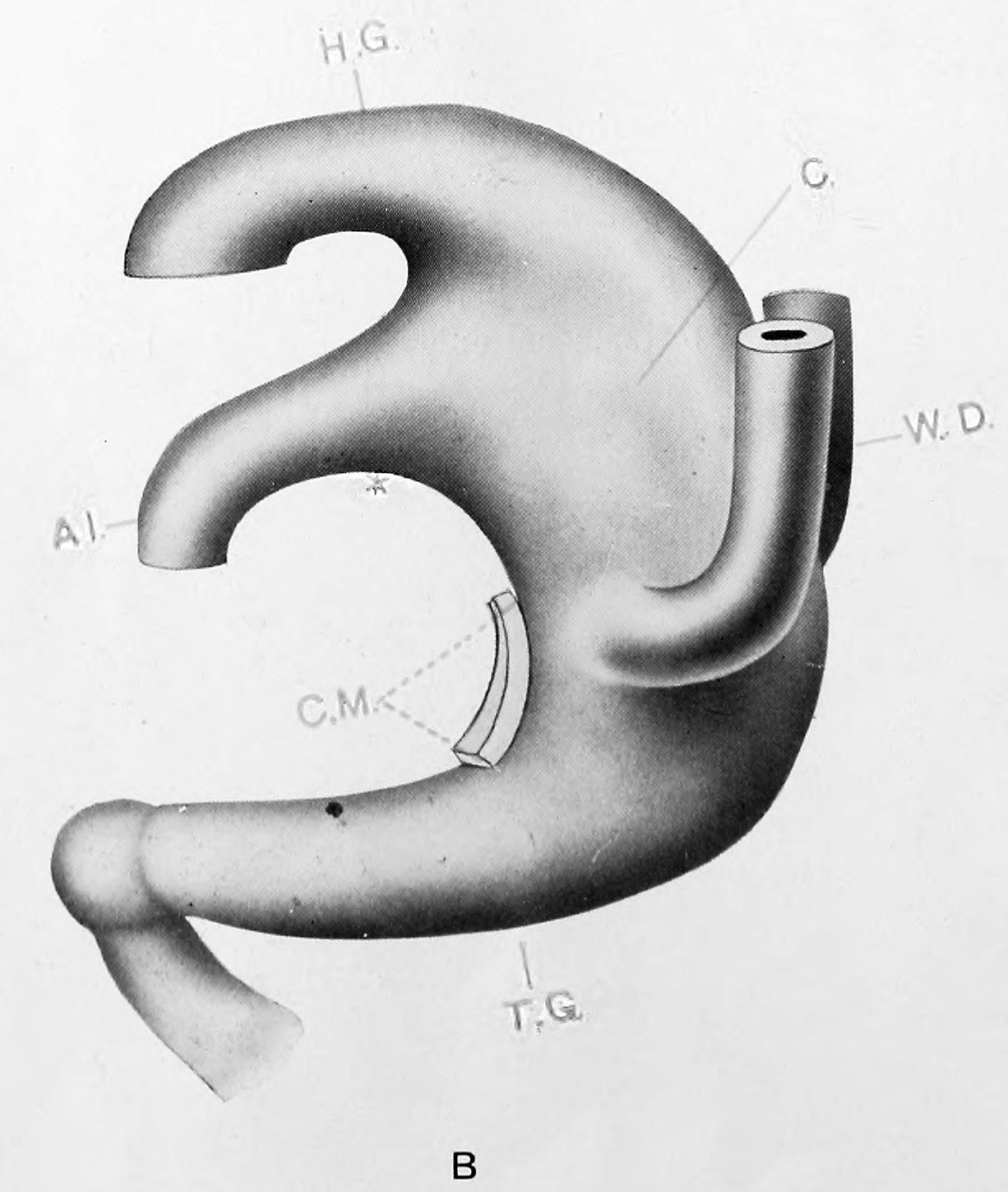
Plate 1B
Model B Embryo No. 5 (Mall 186; 3.5 mm.; +; 20; Good). x 100. Allantois wide lumened and no distinct line of demarkation from the ventral cloacal segment. Wolflian ducts have arrived but not as yet opened. Tail gut maximum of development. Probably the first signs of cloacal division. Carnegie stage 13
Model B {No. 5; Mall no. 186; 3.5 mm.) This model, although made from an embryo of the same length as the above, shows a distinctly older stage in the development of the tract. The allantois has a much wider lumen and the angle between it and the hind gut is much more acute. The Wolffian ducts have reached the cloaca, and attach but do not open into it ventrally near the upper limit of the cloacal membrane. The tail gut is much larger both in length and in thickness, and terminates in an undifferentiated cell mass formed by itself, the chorda and the neural tube (shown as a knob-like ending). The cloaca is wider dorsally than ventrally. A stage similar to the one described was found in two other embryos (nos. 7 and 8), in both of which the allantois was wide at its apparent opening into the cloaca. The possibility of some minor degree of development abnormality suggested itself but on careful study, it was decided that the first evidences of cloacal division were at hand.
Model B, other than a better developed tail gut, a slightly higher position of the Wolffian ducts and a shorter cloacal membrane agrees with Keibel's model of the 4.2 mm. embryo. Keibel's model shows this same evidence of division; a frontal septum between the allantois and hind gut slipping down to a level approaching the upper limit of the cloacal membrane. The writer has found no embryo where the cloacal membrane approaches the level of the dermal navel, and will consider this point later.
| Key to Lettering | ||
|---|---|---|
|
A., Allantois K., Kidney A.M., Position of anal membrane P.T., Precloacal mesodermic tissue C., Cloaca |
R.B., Renal bud CM., Cloacal membrane T.G., Tail cut C.S., Cloacal segment of Wolffian duct U., Ureter |
G.E., Genital eminence U.G., Urogenital sinus H.G., Hind gut U.M., Position of urogenital membrane * Probable position of point where allantois joins cloaca. |
All drawings represent 100 diameters enlargement except of Model F which is 50 diameters.
- Links: plate 1 | 1 A | 1 B | plate 2 | plate 2C | plate 2D | plate 3 | plate 3E | plate 3F | Pohlman 1911
Reference
Pohlman AG. The development of the cloaca in human embryos. (1911) Amer. J Anat. 12: 1-26.
Cite this page: Hill, M.A. (2024, April 27) Embryology Pohlman1911 plate1B.jpg. Retrieved from https://embryology.med.unsw.edu.au/embryology/index.php/File:Pohlman1911_plate1B.jpg
- © Dr Mark Hill 2024, UNSW Embryology ISBN: 978 0 7334 2609 4 - UNSW CRICOS Provider Code No. 00098G
File history
Click on a date/time to view the file as it appeared at that time.
| Date/Time | Thumbnail | Dimensions | User | Comment | |
|---|---|---|---|---|---|
| current | 14:24, 15 June 2016 | | 1,226 × 1,449 (180 KB) | Z8600021 (talk | contribs) | ==Plate 1B== '''Model B''' {No. 5; Mall no. 186; 3.5 mm.) This model, although made from an embryo of the same length as the above, shows a distinctly older stage in the development of the tract. The allantois has a m... |
You cannot overwrite this file.
File usage
The following 2 pages use this file: