File:Placental villi 2.jpg: Difference between revisions
From Embryology
No edit summary |
|||
| Line 5: | Line 5: | ||
:'''Links:''' [[:File:Placental_villi.jpg|Image - First trimester villi overview]] | [[:File:Placental_villi_2.jpg|Image - First trimester villi detail]] | [[Fetal Development]] | [[Placenta Development]] | :'''Links:''' [[:File:Placental_villi.jpg|Image - First trimester villi overview]] | [[:File:Placental_villi_2.jpg|Image - First trimester villi detail]] | | [[:File:Placental_villi_3.jpg|Image - Term villi detail]] | [[Fetal Development]] | [[Placenta Development]] | ||
Revision as of 23:30, 23 August 2011
Human Placental Villi First Trimester
- cross-section image.
- Villi are cover in shell of cytotrophoblast cells.
- Villi core contains mainly mesenchyme cells.
- Links: Image - First trimester villi overview | Image - First trimester villi detail | | Image - Term villi detail | Fetal Development | Placenta Development
placenta, first trimester, human H&E
reproductive system, female, chorionic villi, cytotrophoblast, syncytiotrophoblast
original file name Ple41he.jpg
Image Source: UWA Blue Histology http://www.lab.anhb.uwa.edu.au/mb140/CorePages/FemaleRepro/femalerepro.htm
File history
Click on a date/time to view the file as it appeared at that time.
| Date/Time | Thumbnail | Dimensions | User | Comment | |
|---|---|---|---|---|---|
| current | 02:36, 1 April 2012 | 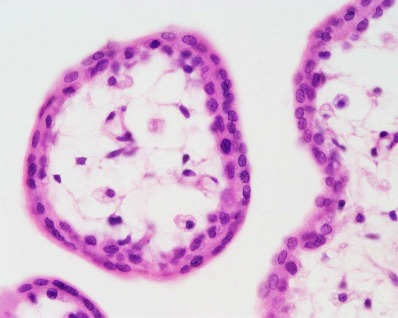 | 1,280 × 1,024 (70 KB) | Z8600021 (talk | contribs) | |
| 16:34, 3 August 2009 | 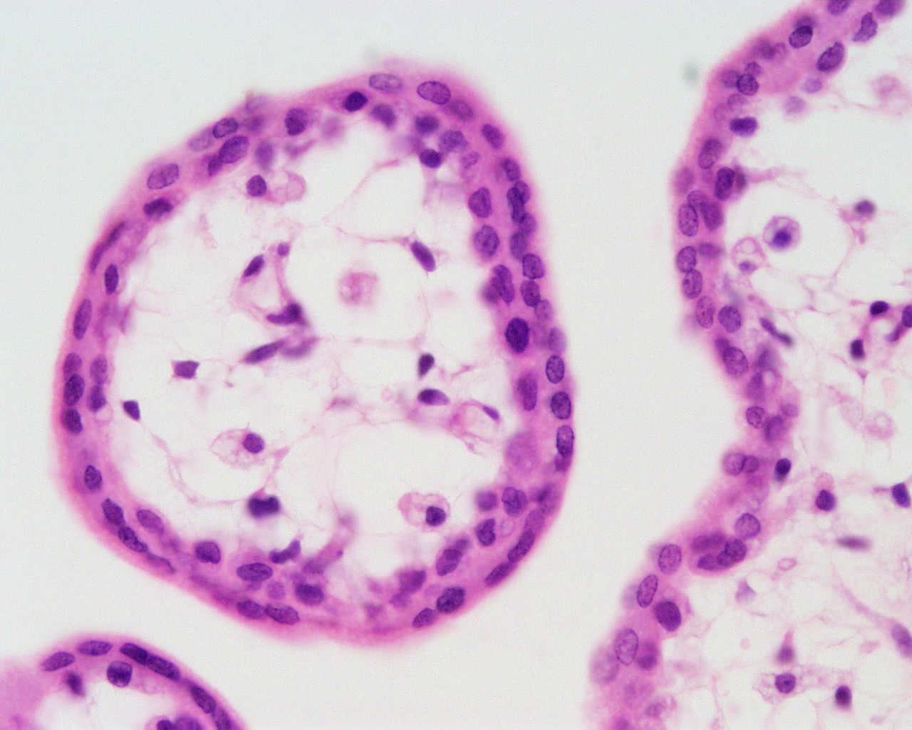 | 1,280 × 1,024 (230 KB) | MarkHill (talk | contribs) | Human placental villi cross-section placenta, first trimester, human H&E reproductive system, female, chorionic villi, cytotrophoblast, syncytiotrophoblast original file name Ple41he.jpg Image Source: UWA Blue Histology http://www.lab.anhb.uwa.edu.au/ |
You cannot overwrite this file.
File usage
The following 4 pages use this file: