File:Ova20he.jpg
From Embryology
No higher resolution available.
Ova20he.jpg (450 × 600 pixels, file size: 96 KB, MIME type: image/jpeg)
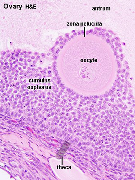
Ovary Histology
- Links: Oocyte Development
Original file name: Ova20he.jpg
Links: Histology | Histology Stains | Blue Histology images copyright Lutz Slomianka 1998-2009. The literary and artistic works on the original Blue Histology website may be reproduced, adapted, published and distributed for non-commercial purposes. See also the page Histology Stains.
Cite this page: Hill, M.A. (2024, May 7) Embryology Ova20he.jpg. Retrieved from https://embryology.med.unsw.edu.au/embryology/index.php/File:Ova20he.jpg
- © Dr Mark Hill 2024, UNSW Embryology ISBN: 978 0 7334 2609 4 - UNSW CRICOS Provider Code No. 00098G
File history
Click on a date/time to view the file as it appeared at that time.
| Date/Time | Thumbnail | Dimensions | User | Comment | |
|---|---|---|---|---|---|
| current | 16:30, 6 May 2012 | | 450 × 600 (96 KB) | Z8600021 (talk | contribs) | |
| 11:16, 28 July 2009 |  | 300 × 400 (59 KB) | MarkHill (talk | contribs) | Ovary Histology Source: UWA Blue Histology |
You cannot overwrite this file.
File usage
The following 25 pages use this file:
- 2009 Lecture 2
- 2010 BGD Lecture - Development of the Embryo/Fetus 1
- 2010 BGD Practical 3 - Oogenesis and Ovulation
- 2010 BGD Practical 3 - Week 3 Summary
- 2010 Lab 2
- 2010 Lecture 2
- 2011 Lab 1 - Oogenesis
- ANAT2241 Female Reproductive System
- ANAT2341 Lab 1 - Oogenesis
- BGDA Lecture - Development of the Embryo/Fetus 1
- BGDA Lecture - Development of the Embryo/Fetus 2
- BGDA Practical - Female Reproductive Tract Histology
- BGDA Practical 3 - Oogenesis and Ovulation
- BGDA Practical 3 - Week 3 Summary
- BGD Lecture - Sexual Differentiation
- Fertilization
- Histology Stains
- Lecture - Fertilization
- Lecture - Genital Development
- Lecture - Week 1 and 2 Development
- Ovary Development
- Timeline human development
- Talk:Timeline human development
- Template:First Trimester Timeline
- Template:First Trimester Timeline collapsable table