File:Mouse-sciatic nerve Schwann cell.jpg
Original file (957 × 1,050 pixels, file size: 339 KB, MIME type: image/jpeg)
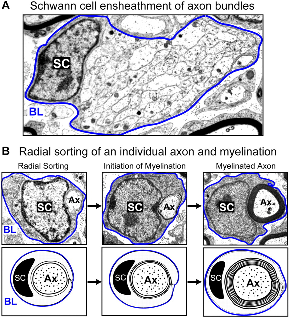
Illustration of Schwann cell (SC) development in the mouse sciatic nerve using electron microscopy. During peripheral nerve development, Schwann cells proliferate, migrate, and ensheath axon (Ax) bundles (A). Schwann cells organize a basal lamina (BL, outlined in blue), which completely surrounds each individual cell and the associated axon(s). Upon receiving the appropriate signals from the local environment, Schwann cells will sort through the axon bundle, isolate an individual axon, and then initiate myelination (B). Bottom panels are schematic representations of the electron micrographs in the top panels, and represent radial sorting, initiation, and myelination of a single axon.
Related myelin images: Mouse - sciatic nerve Schwann cell | sciatic nerve | cerebellum axons | optic nerve
Reference
Chan JR. (2007). Myelination: all about Rac 'n' roll. J. Cell Biol. , 177, 953-5. PMID: 17576794 DOI.
Copyright
This article is distributed under the terms of an Attribution–Noncommercial–Share Alike–No Mirror Sites license for the first six months after the publication date (see http://www.jcb.org/misc/terms.shtml). After six months it is available under a Creative Commons License (Attribution–Noncommercial–Share Alike 3.0 Unported license, as described at http://creativecommons.org/licenses/by-nc-sa/3.0/).
Figure 1. http://jcb.rupress.org/content/177/6/953/F1.large.jpg
Cite this page: Hill, M.A. (2024, April 27) Embryology Mouse-sciatic nerve Schwann cell.jpg. Retrieved from https://embryology.med.unsw.edu.au/embryology/index.php/File:Mouse-sciatic_nerve_Schwann_cell.jpg
- © Dr Mark Hill 2024, UNSW Embryology ISBN: 978 0 7334 2609 4 - UNSW CRICOS Provider Code No. 00098G
File history
Click on a date/time to view the file as it appeared at that time.
| Date/Time | Thumbnail | Dimensions | User | Comment | |
|---|---|---|---|---|---|
| current | 12:50, 24 September 2010 | | 957 × 1,050 (339 KB) | S8600021 (talk | contribs) | Illustration of Schwann cell (SC) development in the mouse sciatic nerve using electron microscopy. During peripheral nerve development, Schwann cells proliferate, migrate, and ensheath axon (Ax) bundles (A). Schwann cells organize a basal lamina (BL, ou |
You cannot overwrite this file.
File usage
The following 2 pages use this file: