File:Mouse-olfactory nerve pathway development.jpg
Original file (600 × 1,764 pixels, file size: 464 KB, MIME type: image/jpeg)
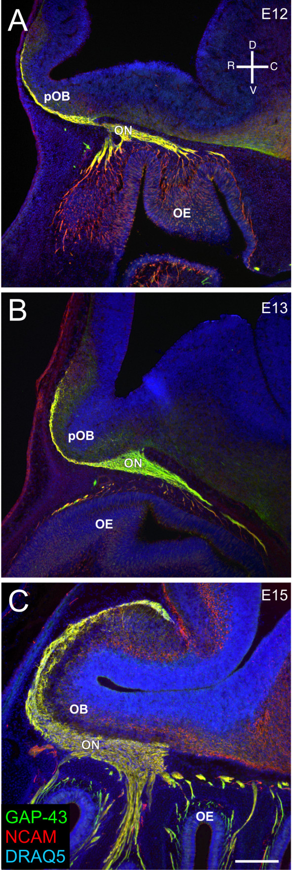
Development of the olfactory nerve pathway
(A-C) Sagittal sections of CD1 embryos stained for GAP-43 (green)/NCAM (red)/DRAQ5 (blue) at E12 (A), E13 (B), and E15 (C). An orientation compass is shown in (A).
Legend
- OE - olfactory epithelium
- ON - olfactory nerve
- pOB - presumptive olfactory bulb
Scale bars = 200 μm.
Miller et al. Neural Development 2010 5:20 doi:10.1186/1749-8104-5-20
Original file name: 1749-8104-5-20-1.jpg
Abstract
- "Olfactory sensory neuron (OSN) axons exit the olfactory epithelium (OE) and extend toward the olfactory bulb (OB) where they coalesce into glomeruli. Each OSN expresses only 1 of approximately 1,200 odor receptors (ORs). OSNs expressing the same OR are distributed in restricted zones of the OE. However, within a zone, the OSNs expressing a specific OR are not contiguous - distribution appears stochastic. Upon reaching the OB the OSN axons expressing the same OR reproducibly coalesce into two to three glomeruli. While ORs appear necessary for appropriate convergence of axons, a variety of adhesion associated molecules and activity-dependent mechanisms are also implicated. Recent data suggest pre-target OSN axon sorting may influence glomerular convergence. Here, using regional and OR-specific markers, we addressed the spatio-temporal properties associated with the onset of homotypic fasciculation in embryonic mice and assessed the degree to which subpopulations of axons remain segregated as they extend toward the nascent OB. We show that immediately upon crossing the basal lamina, axons uniformly turn sharply, usually at an approximately 90° angle toward the OB. Molecularly defined subpopulations of axons show evidence of spatial segregation within the nascent nerve by embryonic day 12, within 48 hours of the first OSN axons crossing the basal lamina, but at least 72 hours before synapse formation in the developing OB. Homotypic fasciculation of OSN axons expressing the same OR appears to be a hierarchical process. While regional segregation occurs in the mesenchyme, the final convergence of OR-specific subpopulations does not occur until the axons reach the inner nerve layer of the OB."
Reference
Miller AM, Maurer LR, Zou DJ, Firestein S & Greer CA. (2010). Axon fasciculation in the developing olfactory nerve. Neural Dev , 5, 20. PMID: 20723208 DOI.
Copyright
© 2010 Miller et al; licensee BioMed Central Ltd. This is an Open Access article distributed under the terms of the Creative Commons Attribution License (http://creativecommons.org/licenses/by/2.0), which permits unrestricted use, distribution, and reproduction in any medium, provided the original work is properly cited.
Cite this page: Hill, M.A. (2024, April 27) Embryology Mouse-olfactory nerve pathway development.jpg. Retrieved from https://embryology.med.unsw.edu.au/embryology/index.php/File:Mouse-olfactory_nerve_pathway_development.jpg
- © Dr Mark Hill 2024, UNSW Embryology ISBN: 978 0 7334 2609 4 - UNSW CRICOS Provider Code No. 00098G
File history
Click on a date/time to view the file as it appeared at that time.
| Date/Time | Thumbnail | Dimensions | User | Comment | |
|---|---|---|---|---|---|
| current | 14:23, 29 September 2010 | 600 × 1,764 (464 KB) | S8600021 (talk | contribs) | ==Development of the olfactory nerve pathway== (A-C) Sagittal sections of CD1 embryos stained for GAP-43 (green)/NCAM (red)/DRAQ5 (blue) at E12 (A), E13 (B), and E15 (C). An orientation compass is shown in (A). Legend * OE - olfactory epithelium * ON |
You cannot overwrite this file.
File usage
The following page uses this file: