File:Mall Meyer1921 table03.jpg

Original file (895 × 1,000 pixels, file size: 289 KB, MIME type: image/jpeg)
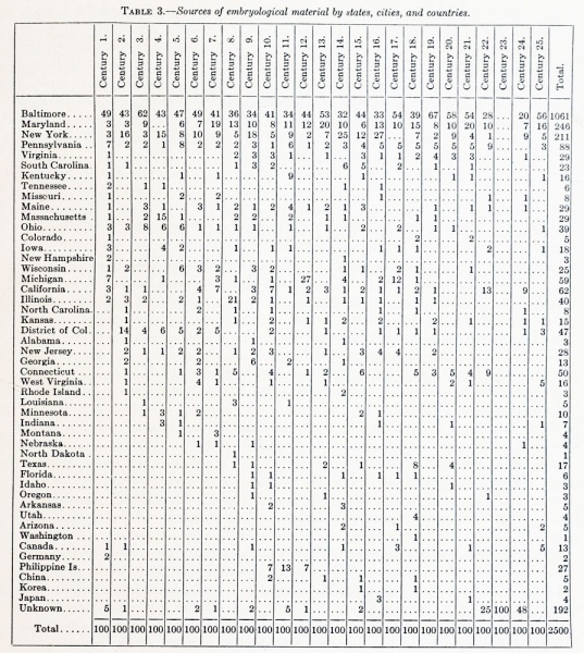
Table 3. Sources of embryological material by states, cities, and countries
Table 3 is interesting as showing the difficulties encountered by embryologists in collecting material. In our own experience we found at first that, although physicians seemed to be entirely willing to send specimens, we rarely received them; or, at best, only those which had been standing upon the shelves for years. These were badly preserved and of little scientific value, not only because the tissues were unfit for microscopic examination, but because histories were entirely lacking. Nevertheless, we always thankfully receive such specimens, and in return gladly send fixing fluids, write letters, and also send reprints of embryological studies to donors. In this way we have learned that a physician who will take the trouble to send one specimen is always willing to preserve carefully the next that falls into his hands, and, in the course of time, he naturally becomes a regular contributor. A number of physicians have been contributors for 30 years, and their unselfish efforts have been a great encouragement to us.
A glance at table 3 will suffice to show that over half of the specimens came from Maryland, and most of these necessarily from Baltimore; next in order is New York, and the third, Pennsylvania. The remainder cover widely scattered points, the first century being drawn from 21 States, the second from 29 States, the sources gradually expanding to include practically every State and Territory of the Union. From the beginning, a few have come regularly from abroad, so that now we have specimens from widely different countries. This wide distribution is due to the systematic circularization of the medical profession. We began by publishing letters of appeal in scientific journals, such as the American Naturalist; but as this was rarely seen by physicians it proved a waste of effort. Then, at the suggestion of Professor Minot, a number of articles were published in Wood's Reference Handbook in the belief that these would come under the eyes of physicians whom we hoped to reach; this plan likewise proved of little value. Finally, a circular was drawn up, widely distributed, and reprinted in most of the medical journals of the country. For a number of years our esteemed colleague, Dr. Howard A. Kelly, inclosed a copy of this circular with every one of his reprints.
| Embryology - 26 Apr 2024 |
|---|
| Google Translate - select your language from the list shown below (this will open a new external page) |
|
العربية | català | 中文 | 中國傳統的 | français | Deutsche | עִברִית | हिंदी | bahasa Indonesia | italiano | 日本語 | 한국어 | မြန်မာ | Pilipino | Polskie | português | ਪੰਜਾਬੀ ਦੇ | Română | русский | Español | Swahili | Svensk | ไทย | Türkçe | اردو | ייִדיש | Tiếng Việt These external translations are automated and may not be accurate. (More? About Translations) |
Mall FP. and Meyer AW. Studies on abortuses: a survey of pathologic ova in the Carnegie Embryological Collection. (1921) Contrib. Embryol., Carnegie Inst. Wash. Publ. 275, 12: 1-364.
- In this historic 1921 pathology paper, figures and plates of abnormal embryos are not suitable for young students.
1921 Carnegie Collection - Abnormal: Preface | 1 Collection origin | 2 Care and utilization | 3 Classification | 4 Pathologic analysis | 5 Size | 6 Sex incidence | 7 Localized anomalies | 8 Hydatiform uterine | 9 Hydatiform tubal | Chapter 10 Alleged superfetation | 11 Ovarian Pregnancy | 12 Lysis and resorption | 13 Postmortem intrauterine | 14 Hofbauer cells | 15 Villi | 16 Villous nodules | 17 Syphilitic changes | 18 Aspects | Bibliography | Figures | Contribution No.56 | Contributions Series | Embryology History
| Historic Disclaimer - information about historic embryology pages |
|---|
| Pages where the terms "Historic" (textbooks, papers, people, recommendations) appear on this site, and sections within pages where this disclaimer appears, indicate that the content and scientific understanding are specific to the time of publication. This means that while some scientific descriptions are still accurate, the terminology and interpretation of the developmental mechanisms reflect the understanding at the time of original publication and those of the preceding periods, these terms, interpretations and recommendations may not reflect our current scientific understanding. (More? Embryology History | Historic Embryology Papers) |
File history
Click on a date/time to view the file as it appeared at that time.
| Date/Time | Thumbnail | Dimensions | User | Comment | |
|---|---|---|---|---|---|
| current | 23:08, 19 November 2012 |  | 895 × 1,000 (289 KB) | Z8600021 (talk | contribs) | ==Table 3. Sources of embryological material by states, cities, and countries== Table 3 is interesting as showing the difficulties encountered by embryologists in collecting material. In our own experience we found at first that, although physicians seem |
You cannot overwrite this file.
File usage
The following 2 pages use this file:
