File:Mall Meyer1921 plate18.jpg

Original file (949 × 1,200 pixels, file size: 220 KB, MIME type: image/jpeg)
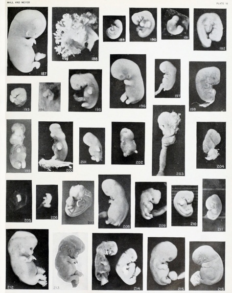
Plate 18
Fig. 187. Slight swelling of fetus from brief maceration. No. 2146. X2.
Fig. 188. Matted, slightly macerated villi. No. 1878. X4.
Fig. 189. A macerated, disproportional cyema 4 mm. long, showing a development of 5.5 mm. No. 786. X4.
Fig. 190. A well-preserved cyema 5.5 mm., of the same development as the preceding. No. 1380. X4.
Figs. 191-192. Cyemata illustrating failure of extension of the body upon maceration. Nos. 1299 ( X2) and 2216 ( X4).
Figs. 193-196. Cyemata illustrating changes in form due to maceration. Nos. 208 (X2), 1296 (X2.67), 1697 (X2), and 1477 (X2).
Fig. 197. Illustrating beginning changes in form due to maceration. No. 705. X1.35.
Figs. 198-204. Similar specimens, showing more pronounced changes. In figure 203 the structure of the specimen is chaotic. Nos. 2244 (X2.67), 1891 (X2), 1655 (X2.67), 1260 (X2.67), 1333 (X2.67), 1379 (X2.67).
Figs. 205-206. Doubtful normally developed cyemata. Nos. 1226 (X2.67) and 2361 (X4).
Fig. 207. A cyema illustrating post-partum changes. No. 589. X2.67.
Fig. 208. Cyema showing maceration sulci and ridges, and drooping of the limbs. No. 52 If. X1.66.
Fig. 209. Cyema showing maceration sulci and ridges, and swelling of the cord. X2.
Fig. 210. Normal, well-preserved cat fetus.
Fig. 211. Normal, poorly preserved cat fetus of approximately the same length.
Fig. 212. Appearance of fetus before fixation. No. 1358. X2.
Fig. 213. The same specimen, showing wrinkling due to fixation and staining. X2.
Fig. 214. Macerated, distorted single-ovum twins. No. 2258. X2.67.
Fig. 215. Minor changes in relief, due to maceration. Swelling and constriction of cord. No. 2014. X2.
Fig. 216. An intermediate maceration form. No. 1495d. X2.67.
| Embryology - 27 Apr 2024 |
|---|
| Google Translate - select your language from the list shown below (this will open a new external page) |
|
العربية | català | 中文 | 中國傳統的 | français | Deutsche | עִברִית | हिंदी | bahasa Indonesia | italiano | 日本語 | 한국어 | မြန်မာ | Pilipino | Polskie | português | ਪੰਜਾਬੀ ਦੇ | Română | русский | Español | Swahili | Svensk | ไทย | Türkçe | اردو | ייִדיש | Tiếng Việt These external translations are automated and may not be accurate. (More? About Translations) |
Mall FP. and Meyer AW. Studies on abortuses: a survey of pathologic ova in the Carnegie Embryological Collection. (1921) Contrib. Embryol., Carnegie Inst. Wash. Publ. 275, 12: 1-364.
- In this historic 1921 pathology paper, figures and plates of abnormal embryos are not suitable for young students.
1921 Carnegie Collection - Abnormal: Preface | 1 Collection origin | 2 Care and utilization | 3 Classification | 4 Pathologic analysis | 5 Size | 6 Sex incidence | 7 Localized anomalies | 8 Hydatiform uterine | 9 Hydatiform tubal | Chapter 10 Alleged superfetation | 11 Ovarian Pregnancy | 12 Lysis and resorption | 13 Postmortem intrauterine | 14 Hofbauer cells | 15 Villi | 16 Villous nodules | 17 Syphilitic changes | 18 Aspects | Bibliography | Figures | Contribution No.56 | Contributions Series | Embryology History
| Historic Disclaimer - information about historic embryology pages |
|---|
| Pages where the terms "Historic" (textbooks, papers, people, recommendations) appear on this site, and sections within pages where this disclaimer appears, indicate that the content and scientific understanding are specific to the time of publication. This means that while some scientific descriptions are still accurate, the terminology and interpretation of the developmental mechanisms reflect the understanding at the time of original publication and those of the preceding periods, these terms, interpretations and recommendations may not reflect our current scientific understanding. (More? Embryology History | Historic Embryology Papers) |
File history
Click on a date/time to view the file as it appeared at that time.
| Date/Time | Thumbnail | Dimensions | User | Comment | |
|---|---|---|---|---|---|
| current | 18:59, 23 November 2012 |  | 949 × 1,200 (220 KB) | Z8600021 (talk | contribs) | ==Plate 18== Fig. 187. Slight swelling of fetus from brief maceration. No. 2146. X2. Fig. 188. Matted, slightly macerated villi. No. 1878. X4. Fig. 189. A macerated, disproportional cyema 4 mm. long, showing a development of 5.5 mm. No. 786. X4. Fig. |
You cannot overwrite this file.
File usage
The following 2 pages use this file:
