File:Mall Meyer1921 fig13.jpg
Original file (1,100 × 800 pixels, file size: 126 KB, MIME type: image/jpeg)
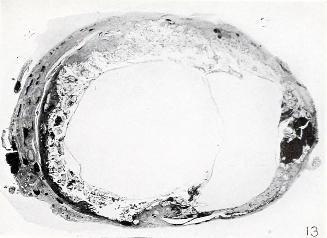
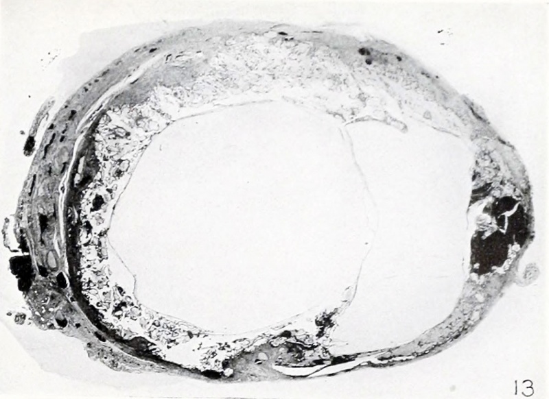
Fig. 13. Hydatiform tubal twins in situ
No. 825. X3.
Many of the tubal specimens are remarkable indeed, and this is true in particular of a case of double-ovum twin pregnancy received from Dr. Cecil Vest. In this specimen the two chorionic vesicles, the intervillous spaces of which were devoid of blood, lay in almost the same transverse diameter of the tube, and hence had distended the latter considerably. Both were implanted quite well over the entire area of contact, which included the whole perimeter of the tube. The chorionic vesicles were flattened at the region of mutual contact, which divided the tube somewhat unequally, as shown in figure 13, one of the original drawings. Although the cyema and the amnion had long disintegrated completely, and although the chorionic membrane itself is thin, covered by degenerate epithelium and also disintegrating, the epithelium of the villi not only is well preserved, but is accompanied by large masses of trophoblast and considerable syncytium. Syncytial buds are found on the chorionic membrane also. The tubal mucosa is largely, and the tubal wall partly, destroyed by the invading trophoblast. Only a few small vestiges of the walls of the villous vessels remain, and the stroma of all of the villi has undergone changes characteristic of hydatiform degeneration. One villus also contains an epithelial cyst resulting from epithelial imagination with subsequent isolation of the distal extremity, a process to be referred to again in connection with the uterine specimens. Since most of these villi still are implanted in the tube, there no longer can be any question as to the conditions under which hydatiform changes in the stroma of the villi are inaugurated. As illustrated in previous instances in which isolated and small groups of villi were still implanted, the advent of degeneration of the stroma usually, if not always, occurs, in part at least, before the villus is detached. Hence it is not merely a maceration change.
- Plate 2: Fig. 12 | Fig. 13 | Fig. 14 | Fig. 15 | Fig. 16 | Fig. 17 | Fig. 18 | Fig. 19 | Chapter 4 Pathologic analysis
| Embryology - 27 Apr 2024 |
|---|
| Google Translate - select your language from the list shown below (this will open a new external page) |
|
العربية | català | 中文 | 中國傳統的 | français | Deutsche | עִברִית | हिंदी | bahasa Indonesia | italiano | 日本語 | 한국어 | မြန်မာ | Pilipino | Polskie | português | ਪੰਜਾਬੀ ਦੇ | Română | русский | Español | Swahili | Svensk | ไทย | Türkçe | اردو | ייִדיש | Tiếng Việt These external translations are automated and may not be accurate. (More? About Translations) |
Mall FP. and Meyer AW. Studies on abortuses: a survey of pathologic ova in the Carnegie Embryological Collection. (1921) Contrib. Embryol., Carnegie Inst. Wash. Publ. 275, 12: 1-364.
- In this historic 1921 pathology paper, figures and plates of abnormal embryos are not suitable for young students.
1921 Carnegie Collection - Abnormal: Preface | 1 Collection origin | 2 Care and utilization | 3 Classification | 4 Pathologic analysis | 5 Size | 6 Sex incidence | 7 Localized anomalies | 8 Hydatiform uterine | 9 Hydatiform tubal | Chapter 10 Alleged superfetation | 11 Ovarian Pregnancy | 12 Lysis and resorption | 13 Postmortem intrauterine | 14 Hofbauer cells | 15 Villi | 16 Villous nodules | 17 Syphilitic changes | 18 Aspects | Bibliography | Figures | Contribution No.56 | Contributions Series | Embryology History
| Historic Disclaimer - information about historic embryology pages |
|---|
 |
File history
Click on a date/time to view the file as it appeared at that time.
| Date/Time | Thumbnail | Dimensions | User | Comment | |
|---|---|---|---|---|---|
| current | 14:27, 24 November 2012 | | 1,100 × 800 (126 KB) | Z8600021 (talk | contribs) | ==Fig. 13. Hydatiform tubal twins in situ== No. 825. X3. :'''Plate 2''': Fig. 12 | Fig. 13 | Fig. 14 | [[:Fil |
You cannot overwrite this file.
File usage
The following 2 pages use this file: