File:Mall Meyer1921 fig11.jpg
Original file (867 × 800 pixels, file size: 118 KB, MIME type: image/jpeg)
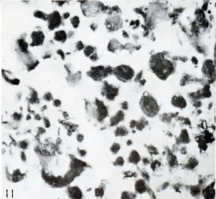
Fig. 11. Almost total lysis or gossamer form
No. 606. X69.
It was especially interesting that this group also contained specimens illustrating the process of intrauterine destruction and absorption of early conceptuses. In one specimen (No. 606) measuring 18 by 13 by 6 mm., all the tissues, even including the last or smallest remnant of the nuclei, had been destroyed completely. Not a single cell contour was preserved; not even by coagulum, as is frequently the case in non-degenerate deciduse. Yet in spite of these things, the form and relative proportions of the entire vesicle, with its surrounding villi, were preserved so well that in describing the gross specimen, Mall noted: "In appearance the specimen is normal," although under later microscopical examination he found it "difficult to make out any structure whatsoever. In fact, even the nuclei of the chorionic membrane have disappeared entirely, leaving only a fine reticular structure." One might say of this specimen, a photograph of some of the villi of which is shown in figure 11, that only an extremely finely textured, disordered web of hyalin material composes the apparently intact chorionic vesicle and the enveloping villi. What we really have here is a cast of the entire chorionic vesicle, including villi, which is formed by hyalin degeneration products that have preserved the form of the vesicle in every detail. From these things it is evident that only a little longer retention of this vesicle in utero would have sufficed to effect its complete disappearance. It does not therefore follow, however, that the disintegration products necessarily would have been completely absorbed. They might, to be sure, have been expelled, at least in part, with the decidua.
The finding of this specimen recalled a personal communication made to me by Professor Mall which suggests that a small cyema received some years ago probably was in a similar structural condition. This specimen was found apparently well-preserved and normal in form when the intact chorionic vesicle was opened in the laboratory, but had completely disintegrated a few moments later.
| Embryology - 27 Apr 2024 |
|---|
| Google Translate - select your language from the list shown below (this will open a new external page) |
|
العربية | català | 中文 | 中國傳統的 | français | Deutsche | עִברִית | हिंदी | bahasa Indonesia | italiano | 日本語 | 한국어 | မြန်မာ | Pilipino | Polskie | português | ਪੰਜਾਬੀ ਦੇ | Română | русский | Español | Swahili | Svensk | ไทย | Türkçe | اردو | ייִדיש | Tiếng Việt These external translations are automated and may not be accurate. (More? About Translations) |
Mall FP. and Meyer AW. Studies on abortuses: a survey of pathologic ova in the Carnegie Embryological Collection. (1921) Contrib. Embryol., Carnegie Inst. Wash. Publ. 275, 12: 1-364.
- In this historic 1921 pathology paper, figures and plates of abnormal embryos are not suitable for young students.
1921 Carnegie Collection - Abnormal: Preface | 1 Collection origin | 2 Care and utilization | 3 Classification | 4 Pathologic analysis | 5 Size | 6 Sex incidence | 7 Localized anomalies | 8 Hydatiform uterine | 9 Hydatiform tubal | Chapter 10 Alleged superfetation | 11 Ovarian Pregnancy | 12 Lysis and resorption | 13 Postmortem intrauterine | 14 Hofbauer cells | 15 Villi | 16 Villous nodules | 17 Syphilitic changes | 18 Aspects | Bibliography | Figures | Contribution No.56 | Contributions Series | Embryology History
| Historic Disclaimer - information about historic embryology pages |
|---|
 |
File history
Click on a date/time to view the file as it appeared at that time.
| Date/Time | Thumbnail | Dimensions | User | Comment | |
|---|---|---|---|---|---|
| current | 13:51, 24 November 2012 | | 867 × 800 (118 KB) | Z8600021 (talk | contribs) | ==Fig. 11. Almost total lysis or gossamer form== No. 606. X69. :'''Plate 1''': Fig 6 | Fig 7 | Fig 8 | [[:Mall_Meyer1921_fig09 |
You cannot overwrite this file.
File usage
The following 2 pages use this file: