File:Macklin-plate05.jpg

Original file (2,331 × 3,061 pixels, file size: 925 KB, MIME type: image/jpeg)
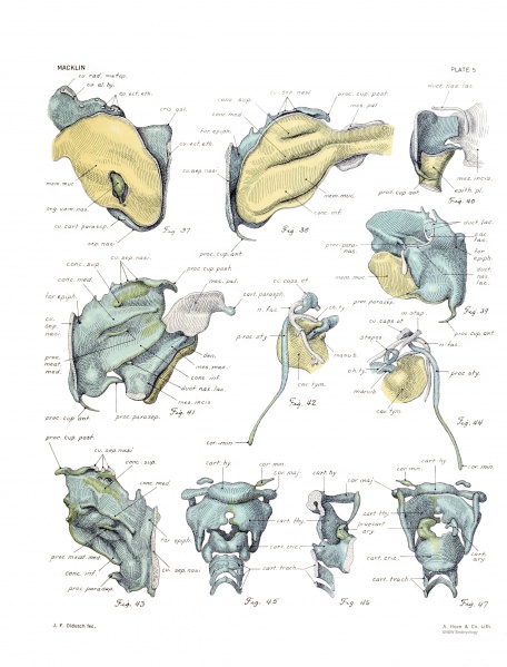
Plate 5. The skull of a human fetus of 43 millimeters greatest length
All drawings were made by Mr. James F. Didusch according to geometric projection. With the exception of figure 7, which was made from a profile reconstruction, all figures were drawn from the original plaster-of-paris models made from human fetus No. 886 of the collection of the Carnegie Laboratory of Embryology. The number of the model from which each figure was drawn is given, together with the magnification. Note - the magnifications refer to the original print versions, not the online images. ReferenceMacklin CC. the skull of a human fetus of 43 millimeters greatest length. (1921) Contrib. Embryol., Carnegie Inst. Wash. Publ., 48, 10:59-102. Cite this page: Hill, M.A. (2024, April 27) Embryology Macklin-plate05.jpg. Retrieved from https://embryology.med.unsw.edu.au/embryology/index.php/File:Macklin-plate05.jpg
In general, blue is used to indicate cartilage and precartilage, yellow to indicate bone, and green for beginning ossification centers. Cut edges are white. The chief departures from this scheme are as follows: Plate 5. — Figures 37, 38, 40, 42, and 44, mucous membrane yellow, precartilage green; in figure 37, epithehum of Jacobson's organ green; in figures 38 and 40, mesenchyme white; in figure 41, teeth yellow, precartilage green, mesenchyme and epithelium of nasolacrimal duct white. Figures 43, 45, 46, and 47, precartilage green. |
Virtual Slide
|
Fig. 37. Mucous membrane of inner wall of right nasal cavity, overlying the septum; it is cut away to show the right organ of Jacobson. (Compare with figure 10.) Model 20. X12.5.
Fig 38. Mucous membrane of lateral wall of right nasal cavity, overljing the right ectethmoid, showing folds for the developing conchce. The mucous membrane fits over the structures seen in figure 41. The elongated nasopharyngeal canal, flanked by the developing palate and medial pterygoid plate, is well seen. Model IS. X12.5.
Fig. 39. Lateral aspect of right ectethmoid from the front, side, and a Uttle below, showing especially the nasolacrimal duct, with the nasolacrimal sac and the lacrimal ducts above, and, below,the expanded end, applied to but not perforating the external aspect of the mucous membrane of the inferior meatus. The tip of the paranasal cartilage hes just lateral to the duct and the tiny shred of osseous tissue representing the lacrimal bone is seen lying along the posterior maxillary process. The cupular process of precartilage is conspicuous in the lower part of the figure. The broad plate of epitheUum, which represents the future inferior meatus, but which has not yet undergone cleavage except posteriorly, is plainly shown. Model 18. X12.5.
Fig. 40. Anterior end of right ectethmoid, with the epithehal plug in the anterior naris, embraced medially by the cupular process of precartilage. The terminal portion of the nasolacrimal duct is shown, entering the space for the inferior meatus, with a small portion of the mesenchyme of the maxilla. Model 19. X12.5.
Fig. 41. Medial aspect of right ectethmoid, showing the developing concha. Precartilage is especially evident in the superior concha, the small process of the middle meatus, and the cupular process. The other conchce are edged with it. Themesenchyme envelopes of the maxilla, the palate, and the medial pterygoid plate are seen. The cartilaginous hamular process is conspicuous, as are also the developing teeth of the right side of the upper jaw. (Compare figure 30.)
Note also the tip of the nasolacrimal duct in the space for the inferior meatus. Model 25. X12.5.
Fig. 42. View of right hyoid arch from without, below, and behind. The connection with the otic capsule is seen above and below appears the lesser cornu of the hyoid cartilage. The relations of the facial nerve, chorda tympani, and tympanic cavity are well seen, and the handle of the malleus is plainly shown in a concavity representing the stratum mucosum of the future tympanic membrane. Model 17. X12.5.
Fig. 43 Medial aspect of left ectethmoid (compare with figure 41), showing developing conchse. The anterior portion of the tectum nasi has been trimmed a little farther laterally than on the right side, and hence the cut surface is not quite the same in the two figures. The cupular process is omitted. Model 3. X12.5.
Fig. 44. View of right hyoid arch from within and slightly above, with its membranous connection with the lesser cornu of the hyoid below (as in fig. 42) and the cut edge of its connection with otic capsule above. Fitting into the curvature of its upper portion is the epithelium of the developing tympanic cavity, which from this point of view is almost parallel with the plane of the paper. The ring-like stapes is seen and to it is attached the tendon of the stapedius muscle, with the muscle itself passing medial to the facial nerve and to the upper end of the styloid process. The handle of the malleus is also seen, with the chorda tympani lying just medial to it. Model 17. X12.5.
Fig. 45. Cartilages of the hyoid, thyroid, cricoid, and upper end of the trachea, seen from the front. On the left side of the model the precartilaginous edging of the upper three cartilages is shown. Model 12. X12.5.
Fig. 46. Right haK of the same structures seen in figures 45 and 47, viewed from within. The precartilage of the arj'tenoid is seen and should be compared with the young cartilage of the same structure shown in figure 47. Model 13. X12.5.
Fig. 47. Same structures seen in figure 45, but viewed from behind. The asymmetry evident in the arytenoids and in other places is due to the fact that on the left side of the model the precartilage was shown, whereas on the right side only the cartilage and young cartilage appear. Model 12. xl2.5.
File history
Click on a date/time to view the file as it appeared at that time.
| Date/Time | Thumbnail | Dimensions | User | Comment | |
|---|---|---|---|---|---|
| current | 15:34, 23 April 2014 |  | 2,331 × 3,061 (925 KB) | Z8600021 (talk | contribs) | |
| 10:45, 16 February 2011 |  | 846 × 1,113 (175 KB) | S8600021 (talk | contribs) | ==Plate 5. The skull of a human fetus of 43 millimeters greatest length== By Charles C. Macklin. (5 plates containing 47 figures) All drawings were made by Mr. James F. Didusch according to geometric projection. With the exception of figure 7, which was |
You cannot overwrite this file.
File usage
The following 2 pages use this file:
