File:Kollmann637.jpg: Difference between revisions
No edit summary |
mNo edit summary |
||
| Line 34: | Line 34: | ||
Kopf ist in die aufrechte Stellung gebracht worden wegen der leichteren | Kopf ist in die aufrechte Stellung gebracht worden wegen der leichteren | ||
Orientierung. | Orientierung. | ||
[[Category:Cranial Nerve]] | |||
Latest revision as of 23:06, 4 September 2014
Fig. 637. The Urspnias folseadea the cranial nerves is shown
Oculomotor, trigeminal, Facial and auditory nerves, glossopharyngeal, vagus and spinal accessory, hypoglossal. Furthermore the origin of the four upper Cervikalnerven. From a human Embryo of 6.9 mm in length. (Age 4 weeks.) (After he Street.)
The neural tube, shows the various departments (see Figures 602 and 605), besides the optic cup and the maze bubbles. The embryonic head is brought into the upright position for ease of orientation.
- This text is a Google translate computer generated translation and may contain many errors.
Images from - Atlas of the Development of Man (Volume 2)
(Handatlas der entwicklungsgeschichte des menschen)
- Kollmann Atlas 2: Gastrointestinal | Respiratory | Urogenital | Cardiovascular | Neural | Integumentary | Smell | Vision | Hearing | Kollmann Atlas 1 | Kollmann Atlas 2 | Julius Kollmann
- Links: Julius Kollman | Atlas Vol.1 | Atlas Vol.2 | Embryology History
| Historic Disclaimer - information about historic embryology pages |
|---|
 |
Reference
Kollmann JKE. Atlas of the Development of Man (Handatlas der entwicklungsgeschichte des menschen). (1907) Vol.1 and Vol. 2. Jena, Gustav Fischer. (1898).
Cite this page: Hill, M.A. (2024, April 27) Embryology Kollmann637.jpg. Retrieved from https://embryology.med.unsw.edu.au/embryology/index.php/File:Kollmann637.jpg
- © Dr Mark Hill 2024, UNSW Embryology ISBN: 978 0 7334 2609 4 - UNSW CRICOS Provider Code No. 00098G
Fig. 637. Der Urspnias der folseadea Kopfnerven ist dargestellt:
Oculomotorius, Trigeminus,
Facialis und Acusticus, Glossopharyngeus, Vagus und Accessorius, Hypoglossus. Femer der Ursprung der vier oberen Cervikalnerven. Von einem menschlichen
Embryo von 6,9 mm Länge. (Alter 4 Wochen.)
(Nach Street er.)
Das Nervenrohr zeigt die verschiedenen Abteilungen (vergl. die Fig. 602 und 605), überdies den Augenbecher und das Labyrinthbläschen. Der embryonale Kopf ist in die aufrechte Stellung gebracht worden wegen der leichteren Orientierung.
File history
Click on a date/time to view the file as it appeared at that time.
| Date/Time | Thumbnail | Dimensions | User | Comment | |
|---|---|---|---|---|---|
| current | 17:26, 17 October 2011 | 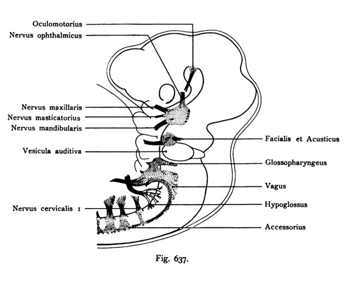 | 679 × 561 (51 KB) | S8600021 (talk | contribs) | Fig. 637. Der Urspnias der folseadea Kopfnerven ist dargestellt: Oculomotorius, Trigeminus, Facialis und Acusticus, Glossopharyngeus, Vagus und Accessorius, Hypoglossus. Femer der Ursprung der vier oberen Cervikalnerven. Von einem menschlichen |
You cannot overwrite this file.
File usage
The following page uses this file: