File:Keibel Mall 2 635.jpg
Original file (1,000 × 1,073 pixels, file size: 223 KB, MIME type: image/jpeg)
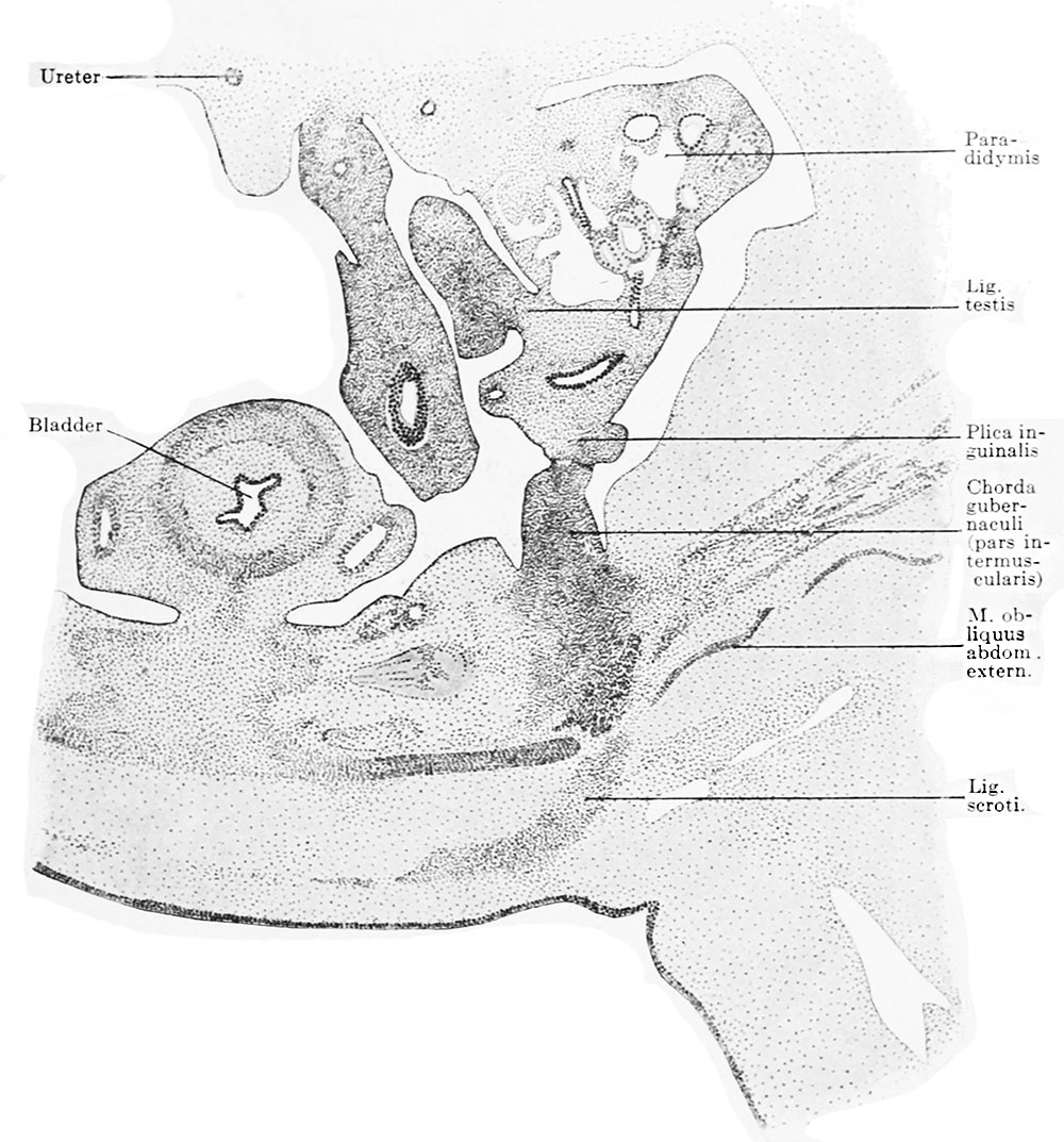
Fig. 635. Portion of a transverse section of a human embryo of 26 mm greatest length
(Embryo R. Meyer 321, from the collection of Professor R. Meyer, Berlin; slide 49, row 5, section 3.) X ca. 30.
The section shows the left mesonephric fold: it has formed a secondary fold, the plica inguinalis. On the posterior surface of the anterior abdominal wall a crista inguinalis has formed, and with it the plica inguinalis has united. The point of union is still distinctly recognizable. Within the crista a cord of compact mesenchyme has appeared, the chorda gubernaculi in the narrower sense. It may be followed through the abdominal wall to an opening in the aponeurosis of the obliquus abdominis externus. From this opening a second cord of compact mesenchyme, the lig. scroti, extends to the integument. The anterior abdominal wall has thickened except at the point of insertion of the crista inguinalis; thus the crista comes to lie in a groove, the anlage of the saccus vaginalis.
| Embryology - 27 Apr 2024 |
|---|
| Google Translate - select your language from the list shown below (this will open a new external page) |
|
العربية | català | 中文 | 中國傳統的 | français | Deutsche | עִברִית | हिंदी | bahasa Indonesia | italiano | 日本語 | 한국어 | မြန်မာ | Pilipino | Polskie | português | ਪੰਜਾਬੀ ਦੇ | Română | русский | Español | Swahili | Svensk | ไทย | Türkçe | اردو | ייִדיש | Tiếng Việt These external translations are automated and may not be accurate. (More? About Translations) |
Felix W. The development of the urinogenital organs. In Keibel F. and Mall FP. Manual of Human Embryology II. (1912) J. B. Lippincott Company, Philadelphia. pp 752-979.
| Historic Disclaimer - information about historic embryology pages |
|---|
 |
Cite this page: Hill, M.A. (2024, April 27) Embryology Keibel Mall 2 635.jpg. Retrieved from https://embryology.med.unsw.edu.au/embryology/index.php/File:Keibel_Mall_2_635.jpg
- © Dr Mark Hill 2024, UNSW Embryology ISBN: 978 0 7334 2609 4 - UNSW CRICOS Provider Code No. 00098G
File history
Click on a date/time to view the file as it appeared at that time.
| Date/Time | Thumbnail | Dimensions | User | Comment | |
|---|---|---|---|---|---|
| current | 09:05, 19 February 2014 | | 1,000 × 1,073 (223 KB) | Z8600021 (talk | contribs) | ==Fig. 635. Portion of a transverse section of a human embryo of 26 mm greatest length== (Embryo R. Meyer 321, from the collection of Professor R. Meyer, Berlin; slide 49, row 5, section 3.) X ca. 30. The section shows the left mesonephric fold: it ... |
You cannot overwrite this file.
File usage
The following 2 pages use this file: