File:Keibel Mall 2 580.jpg

Original file (1,280 × 1,164 pixels, file size: 128 KB, MIME type: image/jpeg)
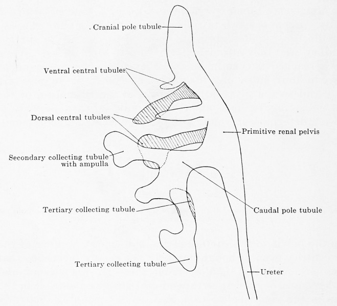
Fig. 580. Ureteric tree of a human embryo of 12.5 mm greatest length
Reconstruction of transverse sections. (Embryo Ma. 1, from the collection of Professor Hochstetter, Vienna.) X 100.
From the primitive renal pelvis six primary collecting tubules have budded out, a cranial and a caudal pole tubule and four central tubules, two ventral and two dorsal. The development of the tubules proceeds in the caudo-cranial direction, the caudal pole tubule has already three secondary tubules, the caudal dorsal central tubule is just dividing, the cranial dorsal one is still pointed, the caudal ventral one is further developed than the cranial ventral one; the cranial pole tubule is still undivided. Of the three secondary tubules from the caudal pole tubule the caudal one has already developed three tertiaries, the cranial is just about to give rise to others, and the middle one has not yet formed an ampulla. Of the primary tubules the cranial pole tubule is a continuation of the ureter or renal pelvis, and the central and caudal pole tubules arise at right angles to the renal pelvis. The cranial secondary tubule is the prolongation of the primary caudal one, the caudal secondary has budded off at right angles. By these different relations of the primary and secondary tubules there is produced the first group-like expansion of the ureteric tree. The dorsal and ventral central tubules form an angle of 45° with one another.
| Embryology - 27 Apr 2024 |
|---|
| Google Translate - select your language from the list shown below (this will open a new external page) |
|
العربية | català | 中文 | 中國傳統的 | français | Deutsche | עִברִית | हिंदी | bahasa Indonesia | italiano | 日本語 | 한국어 | မြန်မာ | Pilipino | Polskie | português | ਪੰਜਾਬੀ ਦੇ | Română | русский | Español | Swahili | Svensk | ไทย | Türkçe | اردو | ייִדיש | Tiếng Việt These external translations are automated and may not be accurate. (More? About Translations) |
Felix W. The development of the urinogenital organs. In Keibel F. and Mall FP. Manual of Human Embryology II. (1912) J. B. Lippincott Company, Philadelphia. pp 752-979.
| Historic Disclaimer - information about historic embryology pages |
|---|
 |
Cite this page: Hill, M.A. (2024, April 27) Embryology Keibel Mall 2 580.jpg. Retrieved from https://embryology.med.unsw.edu.au/embryology/index.php/File:Keibel_Mall_2_580.jpg
- © Dr Mark Hill 2024, UNSW Embryology ISBN: 978 0 7334 2609 4 - UNSW CRICOS Provider Code No. 00098G
File history
Click on a date/time to view the file as it appeared at that time.
| Date/Time | Thumbnail | Dimensions | User | Comment | |
|---|---|---|---|---|---|
| current | 10:14, 13 November 2018 | | 1,280 × 1,164 (128 KB) | Z8600021 (talk | contribs) | |
| 10:09, 13 November 2018 |  | 1,960 × 2,091 (450 KB) | Z8600021 (talk | contribs) |
You cannot overwrite this file.
File usage
The following 2 pages use this file: