File:Keibel Mall 2 566.jpg

Original file (1,280 × 2,311 pixels, file size: 458 KB, MIME type: image/jpeg)
Fig. 566. Model of the mesonephros of a human embryo of 9.5 mm greatest length
(Embryo Ma. 3, from the collection of Professor Hochstetter, Vienna. The model was prepared by my students J. Klausler and Wydler.) Seen from in front. 75.
The two mesonephroi are slightly curved. The individual tubules are pressed together; the Malpighian corpuscles as a result no longer form a single row, but some are beginning to overlap their neighbors. All the tubules are bent into an S-shape. The opening of each tubule into the excretory duct lies about on a level with its Malpighian corpuscle, sometimes slightly more cranially, sometimes slightly more caudally. The various openings are not always the same distance apart. Some tubules are already broken down, on the right the 1st, 3rd and 4th, on the left the 1st, 2nd, 4th and 5th. Some have a common collecting duct, the 15th and 16th, the 19th and 20th. Some Malpighian corpuscles are entirely without tubules, 5a and 27 on the right, 22 on the left. Some tubules are completely formed, but have no opening into the excretory duct, on the right the 16th, on the left the 23rd. The excretory duct is widened in its caudal course from the opening of the 21st tubule, and beyond the opening of the last tubule it narrows again. Seen from in front the bladder appears foreshortened, one looks directly on the cloacal membrane.
| Embryology - 27 Apr 2024 |
|---|
| Google Translate - select your language from the list shown below (this will open a new external page) |
|
العربية | català | 中文 | 中國傳統的 | français | Deutsche | עִברִית | हिंदी | bahasa Indonesia | italiano | 日本語 | 한국어 | မြန်မာ | Pilipino | Polskie | português | ਪੰਜਾਬੀ ਦੇ | Română | русский | Español | Swahili | Svensk | ไทย | Türkçe | اردو | ייִדיש | Tiếng Việt These external translations are automated and may not be accurate. (More? About Translations) |
Felix W. The development of the urinogenital organs. In Keibel F. and Mall FP. Manual of Human Embryology II. (1912) J. B. Lippincott Company, Philadelphia. pp 752-979.
| Historic Disclaimer - information about historic embryology pages |
|---|
 |
Cite this page: Hill, M.A. (2024, April 27) Embryology Keibel Mall 2 566.jpg. Retrieved from https://embryology.med.unsw.edu.au/embryology/index.php/File:Keibel_Mall_2_566.jpg
- © Dr Mark Hill 2024, UNSW Embryology ISBN: 978 0 7334 2609 4 - UNSW CRICOS Provider Code No. 00098G
File history
Click on a date/time to view the file as it appeared at that time.
| Date/Time | Thumbnail | Dimensions | User | Comment | |
|---|---|---|---|---|---|
| current | 07:51, 16 November 2018 | | 1,280 × 2,311 (458 KB) | Z8600021 (talk | contribs) | |
| 07:50, 16 November 2018 | 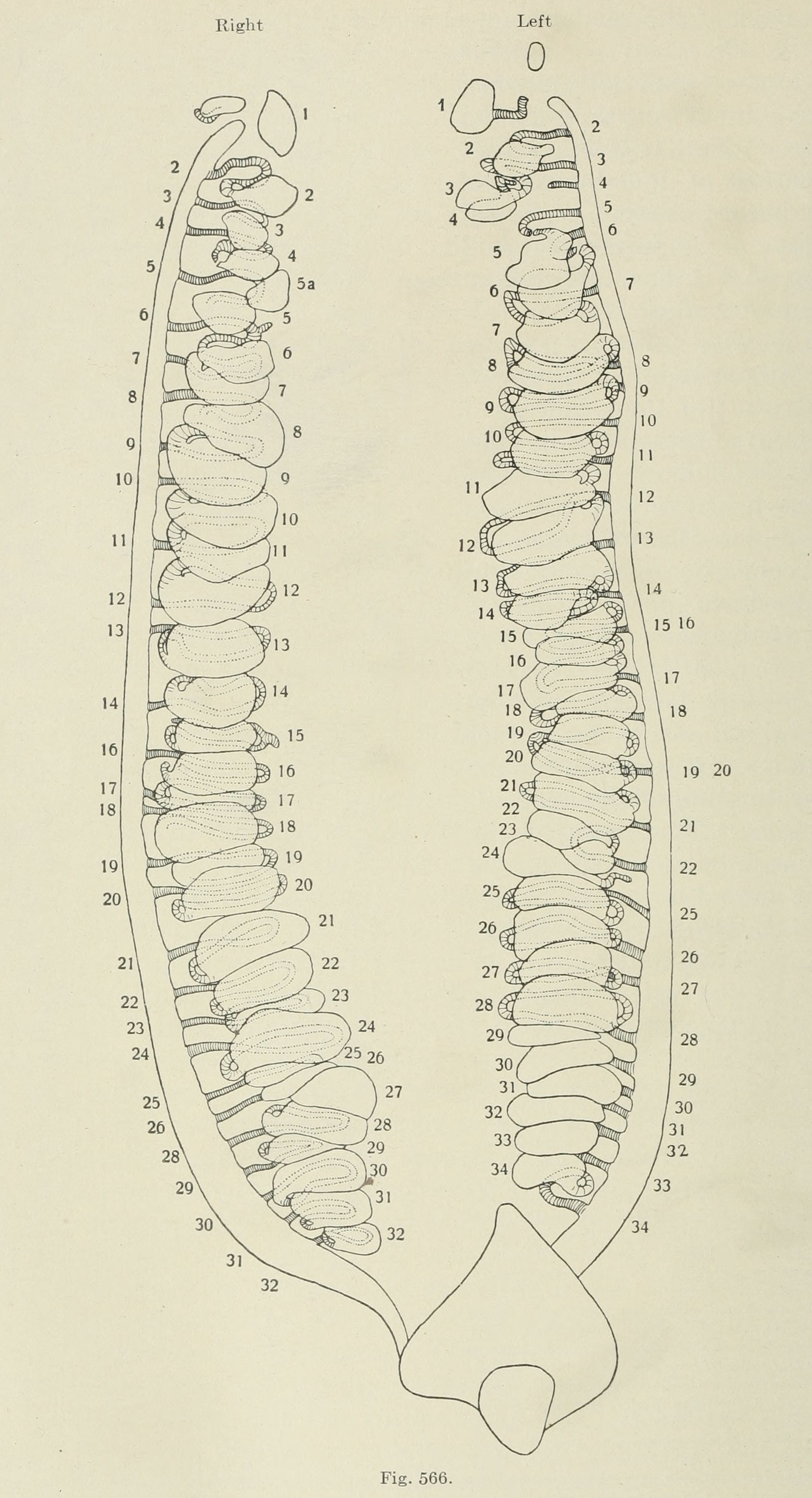 | 1,691 × 3,117 (593 KB) | Z8600021 (talk | contribs) | {{Human Embryology Manual 2 19}} Category:Renal |
You cannot overwrite this file.
File usage
The following 2 pages use this file: