File:Keibel Mall 2 547.jpg
Keibel_Mall_2_547.jpg (648 × 483 pixels, file size: 36 KB, MIME type: image/jpeg)
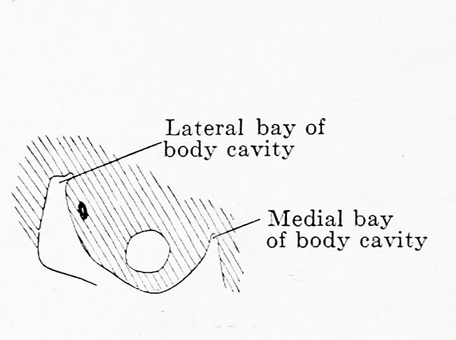
Fig. 547. Transverse section of the middle plate of a human embryo of 4.9 mm nape length
At the level of the 17th primitive segment. (Embryo 139, G. 31, from the collection of the 2nd Anatominal Institute, Berlin [Professor O. Hertwig]. x 150.
The middle plate is evaginated into the body cavity by the development of the mesonephros; it forms the urogenital fold. The fold separates a medial and a lateral bay of the body cavity. In this and the succeeding figures the excretory ducts are black and the Malpighian corpuscles white. These latter lie just below the summit of the urogenital fold, the excretory duct at the ateral surface of its base.
| Embryology - 27 Apr 2024 |
|---|
| Google Translate - select your language from the list shown below (this will open a new external page) |
|
العربية | català | 中文 | 中國傳統的 | français | Deutsche | עִברִית | हिंदी | bahasa Indonesia | italiano | 日本語 | 한국어 | မြန်မာ | Pilipino | Polskie | português | ਪੰਜਾਬੀ ਦੇ | Română | русский | Español | Swahili | Svensk | ไทย | Türkçe | اردو | ייִדיש | Tiếng Việt These external translations are automated and may not be accurate. (More? About Translations) |
Felix W. The development of the urinogenital organs. In Keibel F. and Mall FP. Manual of Human Embryology II. (1912) J. B. Lippincott Company, Philadelphia. pp 752-979.
| Historic Disclaimer - information about historic embryology pages |
|---|
 |
Cite this page: Hill, M.A. (2024, April 27) Embryology Keibel Mall 2 547.jpg. Retrieved from https://embryology.med.unsw.edu.au/embryology/index.php/File:Keibel_Mall_2_547.jpg
- © Dr Mark Hill 2024, UNSW Embryology ISBN: 978 0 7334 2609 4 - UNSW CRICOS Provider Code No. 00098G
File history
Click on a date/time to view the file as it appeared at that time.
| Date/Time | Thumbnail | Dimensions | User | Comment | |
|---|---|---|---|---|---|
| current | 06:25, 15 November 2018 | | 648 × 483 (36 KB) | Z8600021 (talk | contribs) | |
| 06:23, 15 November 2018 | 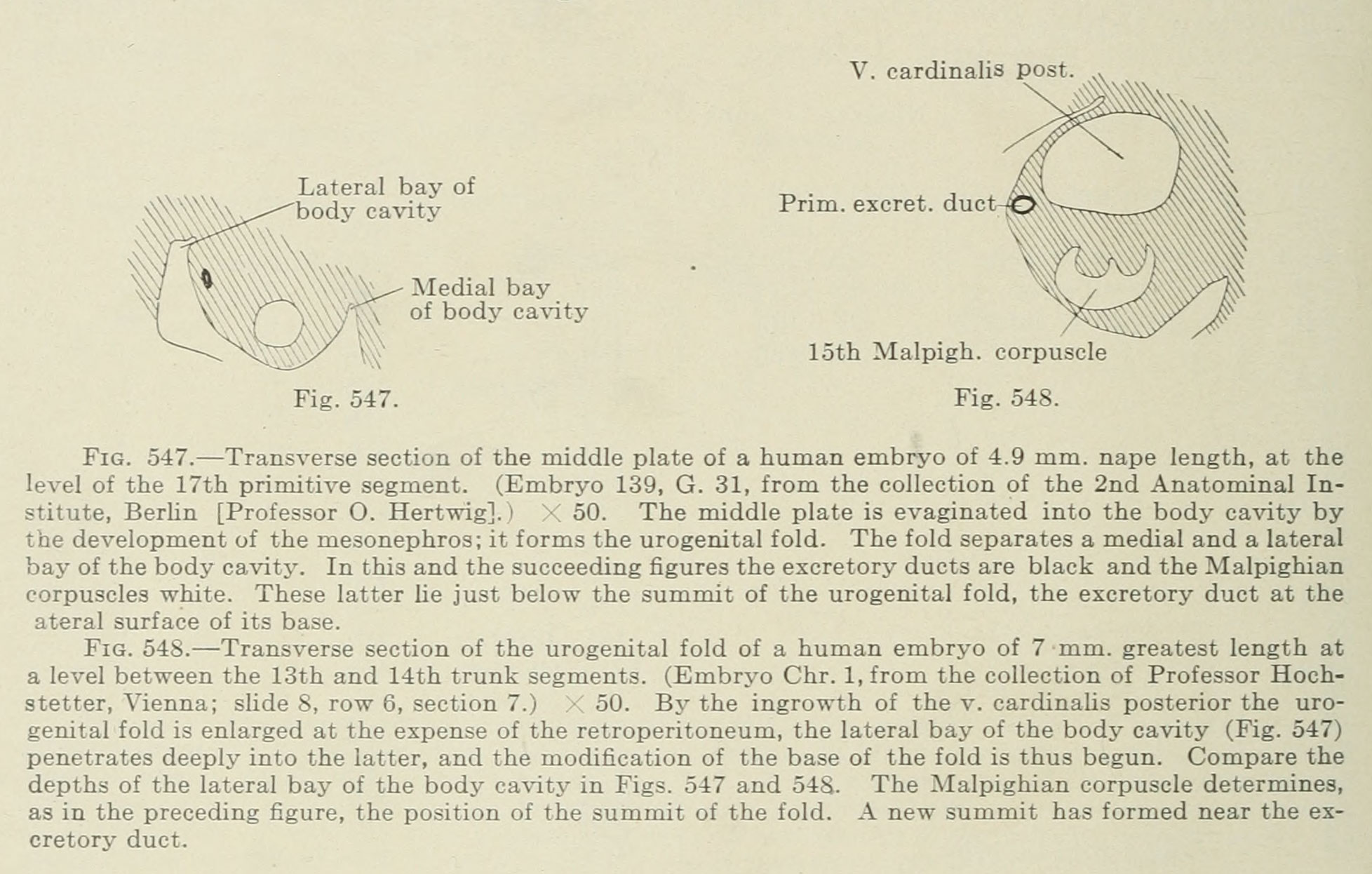 | 1,956 × 1,246 (341 KB) | Z8600021 (talk | contribs) |
You cannot overwrite this file.
File usage
The following 2 pages use this file: