File:Keibel Mall 2 532.jpg
Original file (1,280 × 1,588 pixels, file size: 365 KB, MIME type: image/jpeg)
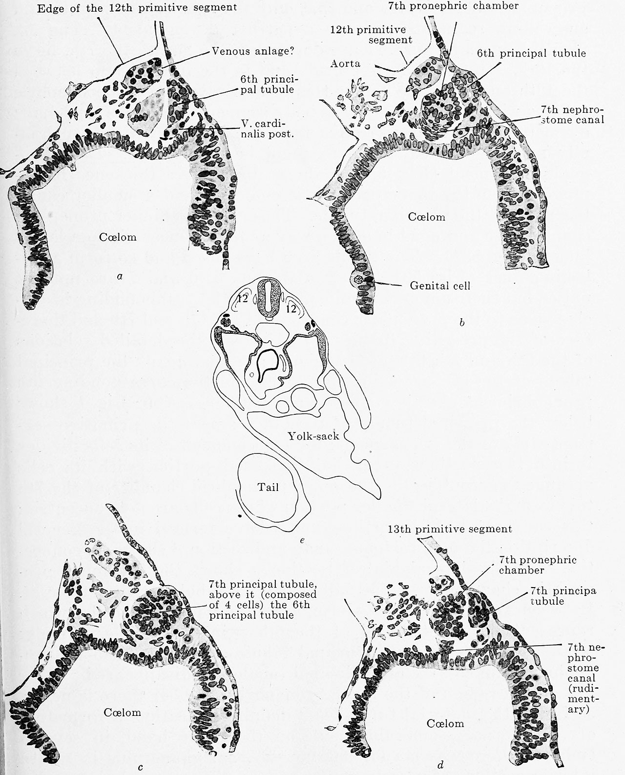
Fig. 532 a, b, c, and d. Four sections from a human embryo of 2.5 mm greatest length
with 23 pairs of primitive segments. (Embryo R. Meyer 300, from the collection of Professor R. Meyer, Berlin; slide 11, row 5, sections 4 and 6; slide 12, row 1, sections 5 and 7.) X 238.
a. Section through the 6th pronephric segment; the caudal wall of the pronephric chamber is just touched, the principal tubule is fully cut. Ventral from the pronephric segment is the anlage of the v. cardinalis post.
b. Section through the 7th pronephric chamber and the 6th principal tubule; in the visceral mesoblast a genital cell.
c. 7th pronephric chamber and 7th principal canal continuous,
d. 7th pronephric segment separated into pronephric chamber and principal canal; rudimentary 7th nephrostome canal. The sections are 5/x thick; if a be taken as the first, then b is the third, c the eighth, and d the tenth section.
| Embryology - 27 Apr 2024 |
|---|
| Google Translate - select your language from the list shown below (this will open a new external page) |
|
العربية | català | 中文 | 中國傳統的 | français | Deutsche | עִברִית | हिंदी | bahasa Indonesia | italiano | 日本語 | 한국어 | မြန်မာ | Pilipino | Polskie | português | ਪੰਜਾਬੀ ਦੇ | Română | русский | Español | Swahili | Svensk | ไทย | Türkçe | اردو | ייִדיש | Tiếng Việt These external translations are automated and may not be accurate. (More? About Translations) |
Felix W. The development of the urinogenital organs. In Keibel F. and Mall FP. Manual of Human Embryology II. (1912) J. B. Lippincott Company, Philadelphia. pp 752-979.
| Historic Disclaimer - information about historic embryology pages |
|---|
 |
Cite this page: Hill, M.A. (2024, April 27) Embryology Keibel Mall 2 532.jpg. Retrieved from https://embryology.med.unsw.edu.au/embryology/index.php/File:Keibel_Mall_2_532.jpg
- © Dr Mark Hill 2024, UNSW Embryology ISBN: 978 0 7334 2609 4 - UNSW CRICOS Provider Code No. 00098G
File history
Click on a date/time to view the file as it appeared at that time.
| Date/Time | Thumbnail | Dimensions | User | Comment | |
|---|---|---|---|---|---|
| current | 17:31, 14 November 2018 | | 1,280 × 1,588 (365 KB) | Z8600021 (talk | contribs) | |
| 17:28, 14 November 2018 |  | 2,038 × 2,973 (761 KB) | Z8600021 (talk | contribs) |
You cannot overwrite this file.
File usage
The following 2 pages use this file: