File:Jenkins003-005.jpg
From Embryology
Size of this preview: 600 × 600 pixels. Other resolution: 1,555 × 1,555 pixels.
Original file (1,555 × 1,555 pixels, file size: 550 KB, MIME type: image/jpeg)
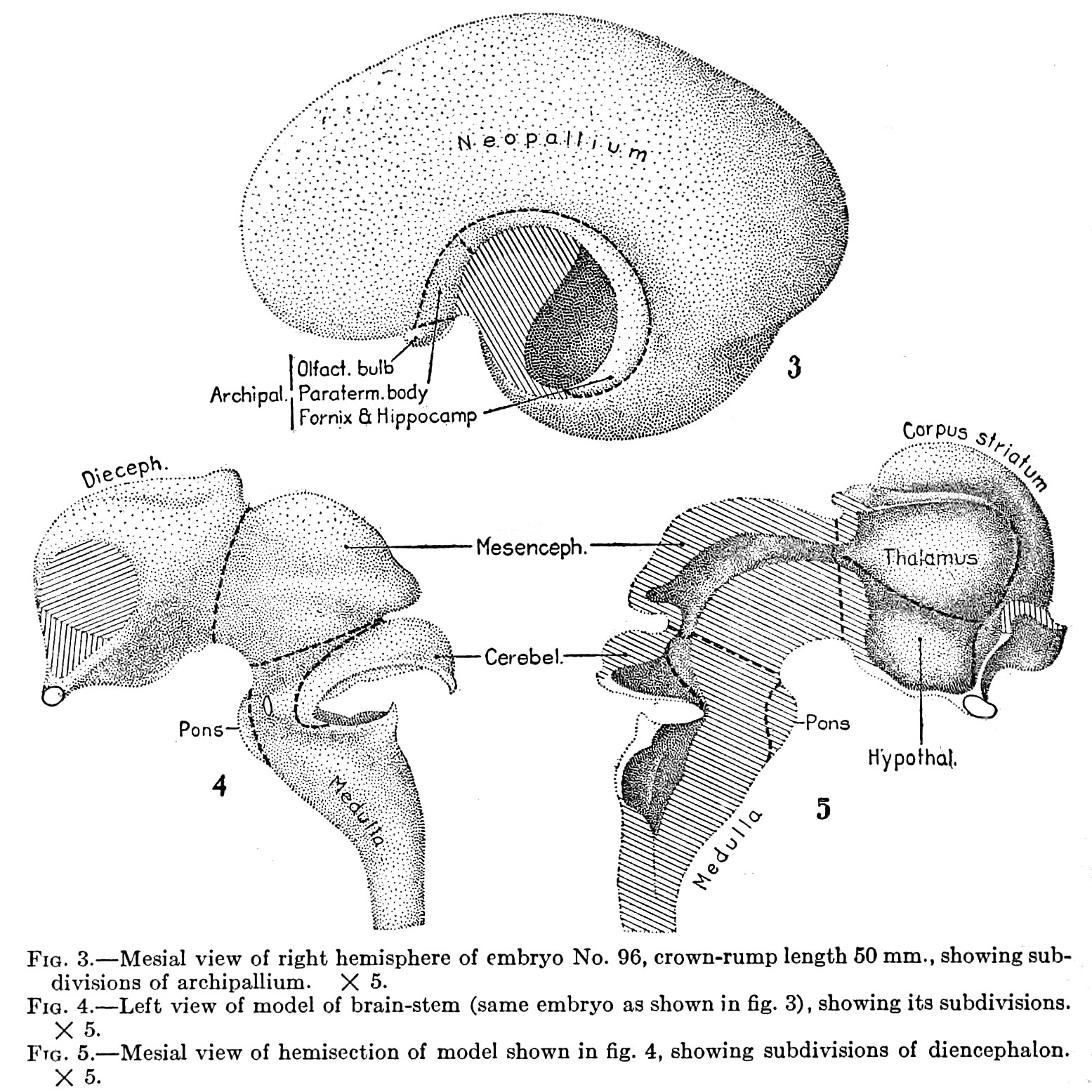
Fig. 3. Mesial view of right hemisphere of embryo
No. 96, crown-rump length 60 mm., showing subdivisions of archipallium. X 5.
Fig. 4. Left view of model of brain-stem
(same embryo as shown in fig. 3), showing its subdivisions. X 5
Fig. 5. Mesial view of hemisection of model shown in fig. 4
showing subdivisions of diencephalon. X 5.
File history
Click on a date/time to view the file as it appeared at that time.
| Date/Time | Thumbnail | Dimensions | User | Comment | |
|---|---|---|---|---|---|
| current | 19:12, 9 March 2015 | | 1,555 × 1,555 (550 KB) | Z8600021 (talk | contribs) | |
| 20:56, 15 February 2011 |  | 745 × 735 (102 KB) | S8600021 (talk | contribs) | ==Fig. 3. Mesial view of right hemisphere of embryo== No. 96, crown-rump length 60 mm., showing subdivisions of archipallium. X 5. ==Fig. 4. Left view of model of brain-stem== (same embryo as shown in fig. 3), showing its subdivisions. X 5 ==Fig. 5. |
You cannot overwrite this file.
File usage
The following page uses this file: