File:Inner ear haircells.jpg: Difference between revisions
From Embryology
| Line 3: | Line 3: | ||
The cochlea and organ of Corti, the sense organ of mammalian hearing. | The cochlea and organ of Corti, the sense organ of mammalian hearing. | ||
(a) The cochlea, a fluid-filled tripartite channel, is located in the inner ear | * (a) The cochlea, a fluid-filled tripartite channel, is located in the inner ear | ||
* (b) A hemisected cochlea provides a radial view of the organ of Corti, a cellular matrix showing the location of hair cells. IHC: inner hair cell OHC: outer hair cell. The input organelles of hair cells, the stereocilia, are connected by different links, including tip-link proteins allowing movement as a unit. | |||
* (c) Deflection of the stereocilary bundle due to displacement between the top of the organ of Corti and the bottom of the tectorial membrane provides tension to the tip link, which, in turn, modulates the MET channel's open probability. | |||
* (d) The tip link is partially composed of cdh23, which is presumed to interact with the MET channel either directly or indirectly. | |||
Legend | |||
*''' Myo1c''' - myosin 1c | |||
* '''CaM''' - calmodulin. | |||
Images in (c) and (d) are modified from LeMasurier and Gillespie PMID 16269359. | |||
===Reference=== | ===Reference=== | ||
<pubmed>19320974</pubmed>| [http://www.ncbi.nlm.nih.gov/pmc/articles/PMC2669096 PMC2669096] | [http://www.biomedcentral.com/1471-2164/10/127 BMC Genomics] | <pubmed>19320974</pubmed>| [http://www.ncbi.nlm.nih.gov/pmc/articles/PMC2669096 PMC2669096] | [http://www.biomedcentral.com/1471-2164/10/127 BMC Genomics] | ||
Revision as of 12:52, 5 October 2011
Anatomical details of inner ear
The cochlea and organ of Corti, the sense organ of mammalian hearing.
- (a) The cochlea, a fluid-filled tripartite channel, is located in the inner ear
- (b) A hemisected cochlea provides a radial view of the organ of Corti, a cellular matrix showing the location of hair cells. IHC: inner hair cell OHC: outer hair cell. The input organelles of hair cells, the stereocilia, are connected by different links, including tip-link proteins allowing movement as a unit.
- (c) Deflection of the stereocilary bundle due to displacement between the top of the organ of Corti and the bottom of the tectorial membrane provides tension to the tip link, which, in turn, modulates the MET channel's open probability.
- (d) The tip link is partially composed of cdh23, which is presumed to interact with the MET channel either directly or indirectly.
Legend
- Myo1c - myosin 1c
- CaM - calmodulin.
Images in (c) and (d) are modified from LeMasurier and Gillespie PMID 16269359.
Reference
<pubmed>19320974</pubmed>| PMC2669096 | BMC Genomics
Copyright © 2009 Zheng et al; licensee BioMed Central Ltd.
This is an Open Access article distributed under the terms of the Creative Commons Attribution License (http://creativecommons.org/licenses/by/2.0), which permits unrestricted use, distribution, and reproduction in any medium, provided the original work is properly cited.
File history
Click on a date/time to view the file as it appeared at that time.
| Date/Time | Thumbnail | Dimensions | User | Comment | |
|---|---|---|---|---|---|
| current | 12:56, 5 October 2011 | 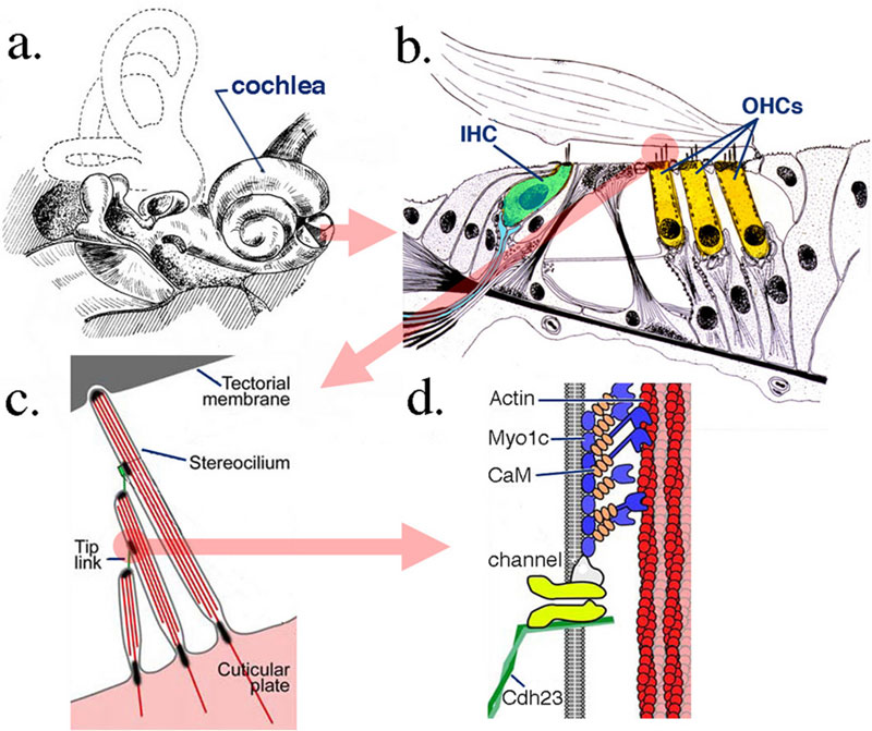 | 800 × 671 (121 KB) | S8600021 (talk | contribs) | |
| 00:59, 28 September 2009 | 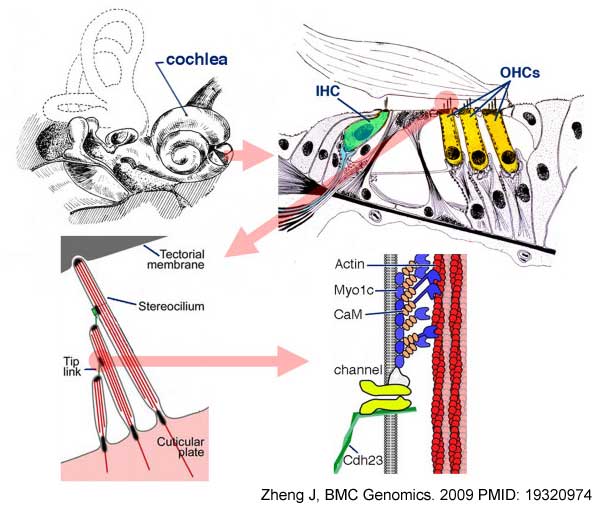 | 600 × 510 (45 KB) | S8600021 (talk | contribs) | Anatomical details of inner ear, cochlea and organ of Corti, the sense organ of mammalian hearing. The cochlea, a fluid-filled tripartite channel, is located in the inner ear (a). A hemisected cochlea provides a radial view of the organ of Corti, a cell |
You cannot overwrite this file.
File usage
The following 6 pages use this file: