File:Hydrocephalus aqueduct of Sylvius 01.jpg

Original file (779 × 726 pixels, file size: 111 KB, MIME type: image/jpeg)
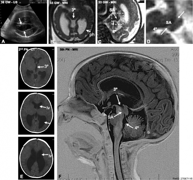
Hydrocephalus - Aqueduct of Sylvius
Progressive obliteration of the aqueduct of Sylvius (SA) in hydrocephalus.
- a An ultrasound of the fetal patient at 36 GW demonstrating dilation of the lateral ventricles (arrows).
- b MRI at 33 GW. The third ventricle (3°) and posterior horns of the lateral ventricles (ph) are dilated.
- c MRI at 33 weeks. Stenosis of the SA is shown.
- d Detailed magnification of the area framed in C showing stenosis of the SA. 4°, fourth ventricle.
- e CT at 39 GW (or the 2nd PN day). The lateral (LV) and third (3°) ventricles are dilated.
- f MRI of the brain on the 5th postnatal day, sagittal T2 imaging. The SA is obliterated
- Links: hydrocephalus | ultrasound | Magnetic Resonance Imaging
Reference
Ortega E, Muñoz RI, Luza N, Guerra F, Guerra M, Vio K, Henzi R, Jaque J, Rodriguez S, McAllister JP & Rodriguez E. (2016). The value of early and comprehensive diagnoses in a human fetus with hydrocephalus and progressive obliteration of the aqueduct of Sylvius: Case Report. BMC Neurol , 16, 45. PMID: 27067115 DOI.
Copyright
This article is distributed under the terms of the Creative Commons Attribution 4.0 International License (http://creativecommons.org/licenses/by/4.0/), which permits unrestricted use, distribution, and reproduction in any medium, provided you give appropriate credit to the original author(s) and the source, provide a link to the Creative Commons license, and indicate if changes were made. The Creative Commons Public Domain Dedication waiver (http://creativecommons.org/publicdomain/zero/1.0/) applies to the data made available in this article, unless otherwise stated.
Fig. 1 12883_2016_566_Fig1_HTML.gif converted to jpg and relabelled.
Cite this page: Hill, M.A. (2024, April 27) Embryology Hydrocephalus aqueduct of Sylvius 01.jpg. Retrieved from https://embryology.med.unsw.edu.au/embryology/index.php/File:Hydrocephalus_aqueduct_of_Sylvius_01.jpg
- © Dr Mark Hill 2024, UNSW Embryology ISBN: 978 0 7334 2609 4 - UNSW CRICOS Provider Code No. 00098G
File history
Click on a date/time to view the file as it appeared at that time.
| Date/Time | Thumbnail | Dimensions | User | Comment | |
|---|---|---|---|---|---|
| current | 18:33, 17 April 2016 |  | 779 × 726 (111 KB) | Z8600021 (talk | contribs) | ==Hydrocephalus - Aqueduct of Sylvius== Progressive obliteration of the aqueduct of Sylvius (SA) in hydrocephalus. a An ultrasound of the fetal patient at 36 GW demonstrating dilation of the lateral ventricles (arrows). b MRI at 33 GW. The third ventr... |
You cannot overwrite this file.
File usage
The following page uses this file: