File:Hydatidiform mole.jpg: Difference between revisions
| Line 2: | Line 2: | ||
Several forms of hydatidiform mole: partial mole, complete mole and persistent gestational trophoblastic tumor. (More? mole types). Many of these tumours arise from a haploid sperm fertilizing an egg without a female pronucleus (the alternative form, an embryo without sperm contribution, is called parthenogenesis). The tumour has a "grape-like" placental appearance without enclosed embryo formation. Following a first molar pregnancy, there is approximately a 1% risk of a second molar pregnancy. | Several forms of hydatidiform mole: partial mole, complete mole and persistent gestational trophoblastic tumor. (More? mole types). Many of these tumours arise from a haploid sperm fertilizing an egg without a female pronucleus (the alternative form, an embryo without sperm contribution, is called parthenogenesis). The tumour has a "grape-like" placental appearance without enclosed embryo formation. Following a first molar pregnancy, there is approximately a 1% risk of a second molar pregnancy. | ||
ACT Pathology Description | ACT Pathology Description | ||
| Line 9: | Line 10: | ||
:'''Links:''' [[Abnormal_Development_-_Hydatidiform_Mole|Hydatidiform Mole]] | [[Placenta_-_Abnormalities|Placenta Abnormalities]] | [[ | :'''Links:''' [[Abnormal_Development_-_Hydatidiform_Mole|Hydatidiform Mole]] | [[Placenta_-_Abnormalities|Placenta Abnormalities]] | [[Placenta Development]] | ||
Revision as of 10:05, 12 May 2013
Hydatidiform Mole
Several forms of hydatidiform mole: partial mole, complete mole and persistent gestational trophoblastic tumor. (More? mole types). Many of these tumours arise from a haploid sperm fertilizing an egg without a female pronucleus (the alternative form, an embryo without sperm contribution, is called parthenogenesis). The tumour has a "grape-like" placental appearance without enclosed embryo formation. Following a first molar pregnancy, there is approximately a 1% risk of a second molar pregnancy.
ACT Pathology Description
- Macroscopic: The specimen is an enlarged uterus (140x140x100mm, weighing 725g) with right and left ovaries and fallopian tubes attached. Opening the uterus shows a cavity filled with a grape-like mass. The wall of the uterus is thickened and fibrous.
- Microscopic: The sections show an interlacing structure within the fibrous lesion. Sections of the cystic contents show numerous dilated and mucoid degenerate proliferative chorionic villi.
Image Source
ACT Pathology, used with permission Professor Julia Potter (Director of Pathology).
ACT Pathology - Uterus benign fibromyoma hydatidiform mole
File history
Click on a date/time to view the file as it appeared at that time.
| Date/Time | Thumbnail | Dimensions | User | Comment | |
|---|---|---|---|---|---|
| current | 21:38, 8 October 2015 | 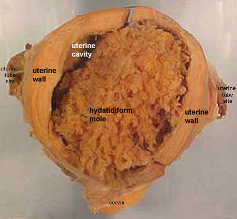 | 800 × 739 (73 KB) | Z8600021 (talk | contribs) | |
| 11:02, 18 August 2009 |  | 400 × 369 (29 KB) | MarkHill (talk | contribs) | Hydatidiform mole Several forms of hydatidiform mole: partial mole, complete mole and persistent gestational trophoblastic tumor. (More? mole types). Many of these tumours arise from a haploid sperm fertilizing an egg without a female pronucleus (the alt |
You cannot overwrite this file.
File usage
The following 23 pages use this file:
- 2009 Lecture 8
- 2010 BGD Practical 3 - Week 2 Summary
- 2010 BGD Tutorial - Applied Embryology and Teratology
- 2010 Lecture 8
- 2011 Lab 2 - Week 2
- ANAT2341 Lab 2 - Week 2
- ANAT2341 Lab 4 - Abnormal Placenta
- ASA Meeting 2013 - Placenta
- Abnormal Development - Hydatidiform Mole
- BGDA Practical 3 - Week 2 Summary
- BGDA Practical Placenta - Abnormalities
- BGD Tutorial - Applied Embryology and Teratology
- Implantation
- Lecture - Fertilization
- Lecture - Placenta Development
- P
- Placenta - Abnormalities
- Placenta Development
- Trophoblast
- Week 2
- Talk:2011 Lab 2 - Week 2
- File:Placenta abnormalities.jpg
- Template:Placenta Abnormalities gallery