File:Human oocyte-metaphase II.jpg: Difference between revisions
No edit summary |
mNo edit summary |
||
| (4 intermediate revisions by 2 users not shown) | |||
| Line 9: | Line 9: | ||
{{Human oocyte to blastocyst}} | [[Cell Division - Meiosis]] | [[Cell Division - Mitosis]] | |||
===Reference=== | |||
http://www.ncbi.nlm.nih.gov/pmc/articles/PMC2773928/ | <pubmed>19924284</pubmed>| [http://www.ncbi.nlm.nih.gov/pmc/articles/PMC2773928 PMC2773928] | [http://www.plosone.org/article/info%3Adoi%2F10.1371%2Fjournal.pone.0007844 PLoS One] | ||
PLoS One. 2009; 4(11): e7844. | |||
Published online 2009 November 16. doi: 10.1371/journal.pone.0007844. | |||
====Copyright==== | |||
Zhang et al. This is an open-access article distributed under the terms of the Creative Commons Attribution License, which permits unrestricted use, distribution, and reproduction in any medium, provided the original author and source are credited. | |||
http://www.ncbi.nlm.nih.gov/pmc/articles/PMC2773928/figure/pone-0007844-g004 | |||
[[Category:Human Embryo]] [[Category:Oocyte]] [[Category:Week 1]] | [[Category:Human Embryo]] [[Category:Oocyte]] [[Category:Week 1]] [[Category:Polar Body]] | ||
Latest revision as of 09:45, 17 June 2014
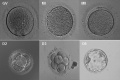
Human Oocyte Morphology
This is a meiosis II metaphase II oocyte, note the absence of an obvious nucleus and the presence of a polar body at the cell periphery. The polar body has pushed the oocyte plasma membrane away from direct contact with the zona pellucida.
Some animal studies suggest that the position of this oocyte plasma membrane/zona pellucida space can determine the eventual site of fertilization.
MII - metaphase II oocyte
Image Links: Human oocyte to blastocyst | Germinal vesicle oocyte (GV) | Metaphase I oocyte | Metaphase II oocyte | Day 2 | Day 3 | Day 5 | Day 5 (label) | Day 5 (colour label)
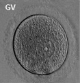
Germinal vesicle oocyte
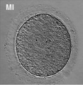
Metaphase I oocyte
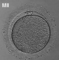
Metaphase II oocyte
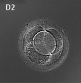
Day 2
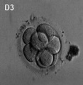
Day 3 - Morula
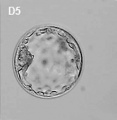
Day 5 - Blastocyst
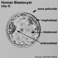
Day 5 (label)
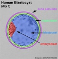
Day 5 (colour label)
Human oocyte to blastocyst


- Links: Oocyte | Morula | Blastocyst | Carnegie stage 1 | Carnegie stage 2 | Carnegie stage 3 | Cell Division - Meiosis | Cell Division - Mitosis
Reference
<pubmed>19924284</pubmed>| PMC2773928 | PLoS One
PLoS One. 2009; 4(11): e7844.
Published online 2009 November 16. doi: 10.1371/journal.pone.0007844.
Copyright
Zhang et al. This is an open-access article distributed under the terms of the Creative Commons Attribution License, which permits unrestricted use, distribution, and reproduction in any medium, provided the original author and source are credited.
http://www.ncbi.nlm.nih.gov/pmc/articles/PMC2773928/figure/pone-0007844-g004
File history
Click on a date/time to view the file as it appeared at that time.
| Date/Time | Thumbnail | Dimensions | User | Comment | |
|---|---|---|---|---|---|
| current | 15:46, 17 April 2012 |  | 400 × 409 (12 KB) | Z8600021 (talk | contribs) | |
| 12:26, 5 April 2010 |  | 320 × 330 (21 KB) | S8600021 (talk | contribs) | Morphology of human oocyte. MII - metaphase II oocyte Note - Original image has been modified to remove array data, colour and other embryo images. See also File:Human-oocyte_to_blastocyst.jpg Pone.0007844.g004.jpg http://www.ncbi.nlm.nih.gov/pm |
You cannot overwrite this file.
File usage
The following 15 pages use this file:
- Cell Division - Meiosis
- F
- In Vitro Oogenesis
- Oocyte Development
- File:Human-oocyte.jpg
- File:Human-oocyte to blastocyst.jpg
- File:Human embryo day 2.jpg
- File:Human embryo day 3.jpg
- File:Human embryo day 5.jpg
- File:Human embryo day 5 label.gif
- File:Human embryo day 5 label.jpg
- File:Human embryo day 5 label2.jpg
- File:Human oocyte-metaphase I.jpg
- File:Human oocyte-metaphase II.jpg
- Template:Human oocyte to blastocyst