File:Human CS13 otic vesicle 01.jpg

Original file (1,028 × 774 pixels, file size: 112 KB, MIME type: image/jpeg)
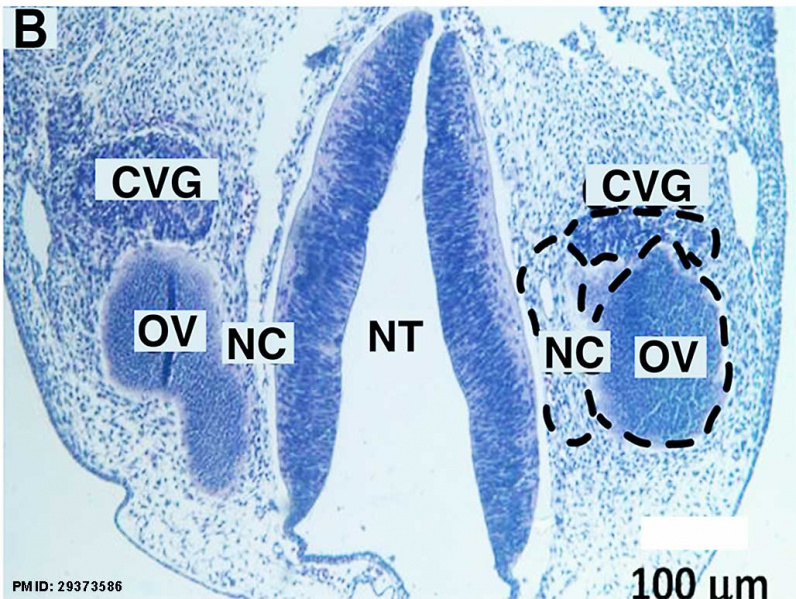
Human Otic Vesicle Carnegie Stages 13 to 15
Laser-microdissection of FFPE-human inner ear slides.
(B) Coronal section of a fetus with highlighted regions of interest (dashed black circles, CVG: cochlear-vestibular ganglions, OV: otic vesicle, NC: neural crest; GG: geniculate ganglion, G-CVG: geniculate- cochleovestibular ganglions, E: epithelium; and NT: neural tube.
Scale bar = 100 μm (B)
Carnegie Stages - 13
Reference
Chadly DM, Best J, Ran C, Bruska M, Woźniak W, Kempisty B, Schwartz M, LaFleur B, Kerns BJ, Kessler JA & Matsuoka AJ. (2018). Developmental profiling of microRNAs in the human embryonic inner ear. PLoS ONE , 13, e0191452. PMID: 29373586 DOI.
Copyright
© 2018 Chadly et al. This is an open access article distributed under the terms of the Creative Commons Attribution License, which permits unrestricted use, distribution, and reproduction in any medium, provided the original author and source are credited.
Fig 1. https://doi.org/10.1371/journal.pone.0191452.g001 Panel B cropped from full figure, adjusted in size and contrast, labelled with PMID.
Cite this page: Hill, M.A. (2024, April 27) Embryology Human CS13 otic vesicle 01.jpg. Retrieved from https://embryology.med.unsw.edu.au/embryology/index.php/File:Human_CS13_otic_vesicle_01.jpg
- © Dr Mark Hill 2024, UNSW Embryology ISBN: 978 0 7334 2609 4 - UNSW CRICOS Provider Code No. 00098G
File history
Click on a date/time to view the file as it appeared at that time.
| Date/Time | Thumbnail | Dimensions | User | Comment | |
|---|---|---|---|---|---|
| current | 12:27, 6 April 2018 |  | 1,028 × 774 (112 KB) | Z8600021 (talk | contribs) |
You cannot overwrite this file.
File usage
There are no pages that use this file.