File:Horseshoe kidney.jpg
From Embryology
No higher resolution available.
Horseshoe_kidney.jpg (776 × 416 pixels, file size: 39 KB, MIME type: image/jpeg)
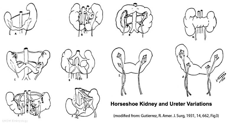
In the horseshoe kidney there is fusion of the lower poles of the kidney.
During migration from the sacral region the two metanephric blastemas can come into contact as shown in Figure 14 mainly at the lower pole. The ureters pass in front of the zone of fusion of the kidneys. The kidneys and ureters usually function adequately but there is an increased incidence of upper urinary tract obstruction or infection.
Some horseshoe variations have been described as having associated ureter abnormalities including duplications.
File history
Click on a date/time to view the file as it appeared at that time.
| Date/Time | Thumbnail | Dimensions | User | Comment | |
|---|---|---|---|---|---|
| current | 13:55, 19 September 2009 | | 776 × 416 (39 KB) | S8600021 (talk | contribs) |
You cannot overwrite this file.