File:Gray0965.jpg

Original file (600 × 750 pixels, file size: 130 KB, MIME type: image/jpeg)
Reflections of the Pleura
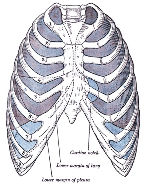
Front view of thorax, showing the relations of the pleuræ and lungs to the chest wall. Pleura in blue; lungs in purple.
Commencing at the sternum, the pleura passes lateralward, lines the inner surfaces of the costal cartilages, ribs, and Intercostales, and at the back part of the thorax passes over the sympathetic trunk and its branches, and is reflected upon the sides of the bodies of the vertebræ, where it is separated by a narrow interval, the posterior mediastinum, from the opposite pleura. From the vertebral column the pleura passes to the side of the pericardium, which it covers to a slight extent; it then covers the back part of the root of the lung, from the lower border of which a triangular sheet descends vertically toward the diaphragm. This sheet is the posterior layer of a wide fold, known as the pulmonary ligament. From the back of the lung root, the pleura may be traced over the costal surface of the lung, the apex and base, and also over the sides of the fissures between the lobes, on to its mediastinal surface and the front part of its root. It is continued from the lower margin of the root as the anterior layer of the pulmonary ligament, and from this it is reflected on to the pericardium (pericardial pleura), and from it to the back of the sternum. Above the level of the root of the lung, however, the mediastinal pleura passes uninterruptedly from the vertebral column to the sternum over the structures in the superior mediastinum. Below, it covers the upper surface of the diaphragm and extends, in front, as low as the costal cartilage of the seventh rib; at the side of the chest, to the lower border of the tenth rib on the left side and to the upper border of the same rib on the right side; and behind, it reaches as low as the twelfth rib, and sometimes even to the transverse process of the first lumbar vertebra. Above, its cupula projects through the superior opening of the thorax into the neck, extending from 2.5 to 5 cm. above the sternal end of the first rib; this portion of the sac is strengthened by a dome-like expansion of fascia (Sibson’s fascia), attached in front to the inner border of the first rib, and behind to the anterior border of the transverse process of the seventh cervical vertebra. This is covered and strengthened by a few spreading muscular fibers derived from the Scaleni.
- Gray's Images: Development | Lymphatic | Neural | Vision | Hearing | Somatosensory | Integumentary | Respiratory | Gastrointestinal | Urogenital | Endocrine | Surface Anatomy | iBook | Historic Disclaimer
| Historic Disclaimer - information about historic embryology pages |
|---|
 |
| iBook - Gray's Embryology | |
|---|---|

|
|
Reference
Gray H. Anatomy of the human body. (1918) Philadelphia: Lea & Febiger.
Cite this page: Hill, M.A. (2024, April 27) Embryology Gray0965.jpg. Retrieved from https://embryology.med.unsw.edu.au/embryology/index.php/File:Gray0965.jpg
- © Dr Mark Hill 2024, UNSW Embryology ISBN: 978 0 7334 2609 4 - UNSW CRICOS Provider Code No. 00098G
File history
Click on a date/time to view the file as it appeared at that time.
| Date/Time | Thumbnail | Dimensions | User | Comment | |
|---|---|---|---|---|---|
| current | 23:24, 21 August 2012 | | 600 × 750 (130 KB) | Z8600021 (talk | contribs) | |
| 10:08, 25 August 2009 |  | 400 × 500 (48 KB) | S8600021 (talk | contribs) | Front view of thorax, showing the relations of the pleuræ and lungs to the chest wall. * pleura - blue * lungs - purple Category:Historic Category:Gray's 1918 Anatomy Category:Respiratory |
You cannot overwrite this file.
File usage
The following 9 pages use this file: