File:Foster007.jpg
Original file (726 × 612 pixels, file size: 68 KB, MIME type: image/jpeg)
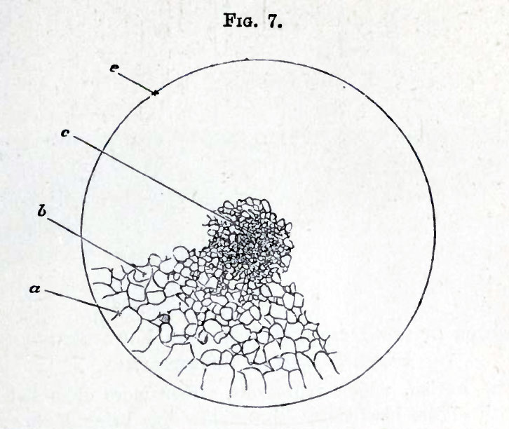
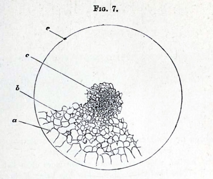
FIG. 7. SURFACE VIEW OF THE GERMINAL Disc OF A HEN'S EGG DURING THE LATER STAGES OF SEGMENTATION.
(Chromic Acid Preparation.)
At c in the centre of the disc the segmentation masses are very small and numerous. At b, nearer the edge, they are larger and fewer ; while those at the extreme margin a are largest and fewest of all. It will be noticed that the radiating furrows marking off the segments a do not reach to the extreme margin e of the disc.
The drawing is completed in one quadrant only ; it will of course be understood that the whole circle ought to be filled up in a precisely similar manner.
| Embryology - 27 Apr 2024 |
|---|
| Google Translate - select your language from the list shown below (this will open a new external page) |
|
العربية | català | 中文 | 中國傳統的 | français | Deutsche | עִברִית | हिंदी | bahasa Indonesia | italiano | 日本語 | 한국어 | မြန်မာ | Pilipino | Polskie | português | ਪੰਜਾਬੀ ਦੇ | Română | русский | Español | Swahili | Svensk | ไทย | Türkçe | اردو | ייִדיש | Tiếng Việt These external translations are automated and may not be accurate. (More? About Translations) |
Foster M. Balfour FM. Sedgwick A. and Heape W. The Elements of Embryology (1883) Vol. 1. (2nd ed.). London: Macmillan and Co.
| Historic Disclaimer - information about historic embryology pages |
|---|
 |
The Elements of Embryology - Volume 1 (1883)
The History of the Chick: Egg structure and incubation beginning | Summary whole incubation | First day | Second day - first half | Second day - second half | Third day | Fourth day | Fifth day | Sixth day to incubation end | Appendix
| Historic Disclaimer - information about historic embryology pages |
|---|
|
Glossary Links
- Glossary: A | B | C | D | E | F | G | H | I | J | K | L | M | N | O | P | Q | R | S | T | U | V | W | X | Y | Z | Numbers | Symbols | Term Link
Cite this page: Hill, M.A. (2024, April 27) Embryology Foster007.jpg. Retrieved from https://embryology.med.unsw.edu.au/embryology/index.php/File:Foster007.jpg
- © Dr Mark Hill 2024, UNSW Embryology ISBN: 978 0 7334 2609 4 - UNSW CRICOS Provider Code No. 00098G
File history
Click on a date/time to view the file as it appeared at that time.
| Date/Time | Thumbnail | Dimensions | User | Comment | |
|---|---|---|---|---|---|
| current | 15:37, 8 January 2011 | | 726 × 612 (68 KB) | S8600021 (talk | contribs) | FIG. 7. SURFACE VIEW OF THE GERMINAL Disc OF A HEN'S EGG DURING THE LATER STAGES OF SEGMENTATION. (Chromic Acid Preparation.) {{Template:Foster 1883}} {{Template:1_Foster_1883}} |
You cannot overwrite this file.
File usage
The following 2 pages use this file: