File:Fetal head medial.jpg: Difference between revisions
No edit summary |
No edit summary |
||
| Line 9: | Line 9: | ||
Original File Name: Headbone2.jpg | Original File Name: Headbone2.jpg | ||
Source: | Source: Prof Virginia Diewert | ||
{{Template:Footer}} | {{Template:Footer}} | ||
[[Category:Human Fetus]] [[Category:Musculoskeletal]] [[Category:Bone]] [[Category:Cartilage]] [[Category:Head]] [[Category:Week 12]] [[Category:Skull]] | [[Category:Human Fetus]] [[Category:Musculoskeletal]] [[Category:Bone]] [[Category:Cartilage]] [[Category:Head]] [[Category:Week 12]] [[Category:Skull]] | ||
Revision as of 12:36, 24 May 2011
Fetal Head (12 weeks)
medial view, stained for bone and cartilage
In this medial (internal) view, note the distribution of cartilage from the nasal region through the base of the skull to the atlas/axis (with new bone forming). See also the original Meckel's cartilage within the newly forming bony mandible.
Note: Cartilage in the neck where the hyoid bone will eventually form. Meckel's cartilage visible in the medial view within the newly forming bone.
Original File Name: Headbone2.jpg
Source: Prof Virginia Diewert
Cite this page: Hill, M.A. (2024, May 22) Embryology Fetal head medial.jpg. Retrieved from https://embryology.med.unsw.edu.au/embryology/index.php/File:Fetal_head_medial.jpg
- © Dr Mark Hill 2024, UNSW Embryology ISBN: 978 0 7334 2609 4 - UNSW CRICOS Provider Code No. 00098G
File history
Click on a date/time to view the file as it appeared at that time.
| Date/Time | Thumbnail | Dimensions | User | Comment | |
|---|---|---|---|---|---|
| current | 15:45, 13 September 2009 | 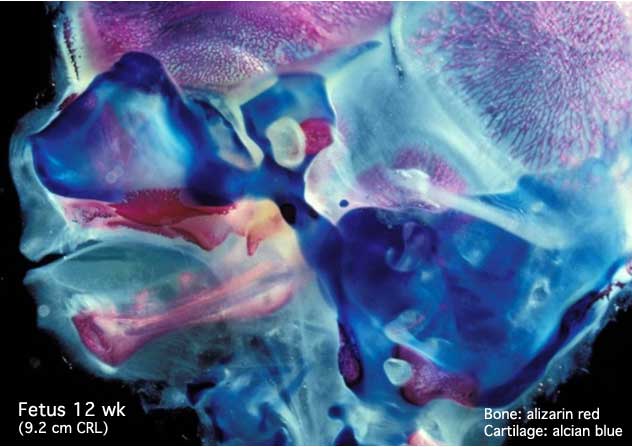 | 632 × 447 (34 KB) | S8600021 (talk | contribs) |
You cannot overwrite this file.
File usage
The following 40 pages use this file:
- 2009 Lecture 13
- 2010 BGD Lecture - Development of the Embryo/Fetus 2
- 2010 BGD Practical 12 - Second Trimester
- 2010 BGD Practical 6 - Week 7
- 2010 Foundations Lecture - Introduction to Human Development
- 2010 Lab 6
- 2010 Lecture 13
- 2011 Lab 12 - Second Trimester
- 2011 Lab 6 - Fetal
- AACP Meeting 2013 - Face Embryology
- ANAT2241 Bone, Bone Formation and Joints
- ANAT2341 Lab 11 - Embryo to Fetus
- ANAT2341 Lab 12 - Second Trimester
- ANAT2341 Lab 6 - Fetal
- Abnormal Development - Fetal Growth Restriction
- BGDA Lecture - Development of the Embryo/Fetus 2
- BGDA Practical 12 - Second Trimester
- BGDA Practical 7 - Week 7
- BGDB Face and Ear - Fetal
- BGD Lecture - Face and Ear Development
- Bone Development
- Bone Histology
- Cartilage Histology
- Fetal Development
- Fetal Development - 12 Weeks
- Foundations Lecture - Introduction to Human Development
- Head Development
- Joint Development - Temporomandibular Joint
- Lecture - Fetal Development
- Lecture - Head Development
- Lecture - Musculoskeletal Development
- Musculoskeletal System - Abnormalities
- Musculoskeletal System - Axial Skeleton Development
- Musculoskeletal System - Bone Development
- Musculoskeletal System - Cartilage Development
- Musculoskeletal System - Joint Development
- Musculoskeletal System - Skull Development
- Musculoskeletal System Development
- Pre-Medicine Program - Embryology
- Virginia Diewert