File:Endochondral ossification 2.jpg: Difference between revisions
No edit summary |
No edit summary |
||
| Line 1: | Line 1: | ||
Endochondral | Endochondral Ossification | ||
Original Image name: Epl04he.jpg | Original Image name: Epl04he.jpg | ||
| Line 6: | Line 6: | ||
Image and Text Source: UWA Blue Histology http://www.lab.anhb.uwa.edu.au/mb140/CorePages/Bone/Bone.htm | Image and Text Source: UWA Blue Histology http://www.lab.anhb.uwa.edu.au/mb140/CorePages/Bone/Bone.htm | ||
{{Template:Blue Histology}} | |||
[[Category:Musculoskeletal]] [[Category:Histology]] | [[Category:Musculoskeletal]] [[Category:Histology]] | ||
Revision as of 11:10, 3 April 2010
Endochondral Ossification
Original Image name: Epl04he.jpg
Image and Text Source: UWA Blue Histology http://www.lab.anhb.uwa.edu.au/mb140/CorePages/Bone/Bone.htm
Links: Histology | Histology Stains | Blue Histology images copyright Lutz Slomianka 1998-2009. The literary and artistic works on the original Blue Histology website may be reproduced, adapted, published and distributed for non-commercial purposes. See also the page Histology Stains.
Cite this page: Hill, M.A. (2024, May 22) Embryology Endochondral ossification 2.jpg. Retrieved from https://embryology.med.unsw.edu.au/embryology/index.php/File:Endochondral_ossification_2.jpg
- © Dr Mark Hill 2024, UNSW Embryology ISBN: 978 0 7334 2609 4 - UNSW CRICOS Provider Code No. 00098G
File history
Click on a date/time to view the file as it appeared at that time.
| Date/Time | Thumbnail | Dimensions | User | Comment | |
|---|---|---|---|---|---|
| current | 13:08, 5 October 2011 | 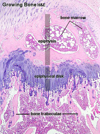 | 400 × 533 (99 KB) | S8600021 (talk | contribs) | increase size of image |
| 11:34, 11 September 2009 |  | 300 × 400 (74 KB) | S8600021 (talk | contribs) | Epl04he.jpg |
You cannot overwrite this file.
File usage
The following 10 pages use this file: